Rothschild, B. M. et al. Recognition of leukemia in skeletal remains: Report and comparison of two cases. Am. J. Phys. Anthropol. Off. Publ. Am. Assoc. Phys. Anthropol. 102, 481–496 (1997).
Rothschild, B. M. Radiologic assessment of osteoarthritis in dinosaurs. Ann. Carnegie Museum 49, 295–301 (1990).
Rothschild, B. M. Diffuse idiopathic skeletal hyperostosis as reflected in the paleontologic record: dinosaurs and early mammals. In Seminars in Arthritis and Rheumatism 17, 119–125 (Elsevier, 1987).
Rothschild, B. M., Witzke, B. J. & Hershkovitz, I. Metastatic cancer in the Jurassic. Lancet 354, 398 (1999).
Bennet, S. C. Pathologies of the large Pterodactyloid pterosaurs Ornithocheirus and Pteranodon. J. Vert. Paleont. 9, 13 (1989).
Tanke, D. H. & Rothschild, B. M. DINOSORES: An Annotated Bibliography of Dinosaur Paleopathology and Related Topics—1838-2001: Bulletin 20. 20 (New Mexico Museum of Natural History and Science, 2002).
Tanke, D. H. & Rothschild, B. M. Paleopathology in Late Cretaceous Hadrosauridae from Alberta, Canada with comments on a putative Tyrannosaurus bite injury on an Edmontosaurus tail. In Hadrosaurs (eds. Eberth, D. & Evans, D.) 540–571 (Bloomington: Indiana University Press, 2014).
Straight, W. H. et al. Bone lesions in hadrosaurs: Computed Tomographic Imaging as a guide for paleohistologic and stable-isotopic analysis. J. Vertebr. Paleontol. 29, 315–325 (2009).
Tumarkin, A. R., Dodson, P., Tanke, D. H. & Rothschild, B. M. Paleohistopathology? A preliminary consider ation of modern fracture repair for interpreting dinosaurian thermophysiology. In Geological Society of America 31, A73 (1999).
Favara, B. E. et al. Contemporary classification of histiocytic disorders. Med. Pediatr. Oncol. Off. J. SIOP—International Soc. Pediatr. Oncol. Societé Int. d’Oncologie Pédiatrique 29, 157–166 (1997).
Huang, W. D. et al. Langerhans cell histiocytosis of spine: A comparative study of clinical, imaging features, and diagnosis in children, adolescents, and adults. Spine J. 13, 1108–1117 (2013).
Allen, C. E., Merad, M. & McClain, K. L. Langerhans-cell histiocytosis. N. Engl. J. Med. 379, 856–868 (2018).
Hoover, K. B., Rosenthal, D. I. & Mankin, H. Langerhans cell histiocytosis. Skeletal Radiol. 36, 95–104 (2007).
Arceci, R. J. The histiocytoses: the fall of the Tower of Babel. Eur. J. Cancer 35, 747–767 (1999).
Berres, M.-L. et al. BRAF-V600E expression in precursor versus differentiated dendritic cells defines clinically distinct LCH risk groups. J. Exp. Med. 211, 669–683 (2014).
Katz, S. I., Tamaki, K. & Sachs, D. H. Epidermal Langerhans cells are derived from cells originating in bone marrow. Nature 282, 324 (1979).
Kiertscher, S. M., Luo, J., Dubinett, S. M. & Roth, M. D. Tumors promote altered maturation and early apoptosis of monocyte-derived dendritic cells. J. Immunol. 164, 1269–1276 (2000).
Ghanem, I., Tolo, V. T., D’Ambra, P. & Malogalowkin, M. H. Langerhans cell histiocytosis of bone in children and adolescents. J. Pediatr. Orthop. 23, 124–130 (2003).
Howarth, D. M. et al. Langerhans cell histiocytosis: diagnosis, natural history, management, and outcome. Cancer 85, 2278–2290 (1999).
Veyssier-Belot, C. et al. Erdheim-Chester disease. Clinical and radiologic characteristics of 59 cases. Medicine (Baltimore). 75, 157–169 (1996).
Haroche, J. et al. Erdheim–Chester disease. Curr. Rheumatol. Rep. 16, 412 (2014).
Bertram, C., Madert, J. & Eggers, C. Eosinophilic granuloma of the cervical spine. Spine (Phila. Pa. 1976). 27, 1408–1413 (2002).
Azouz, E. M., Saigal, G., Rodriguez, M. M. & Podda, A. Langerhans’ cell histiocytosis: pathology, imaging and treatment of skeletal involvement. Pediatr. Radiol. 35, 103–115 (2005).
Meyer, K. A., Bancroft, L. W., Dietrich, T. J., Kransdorf, M. J. & Peterson, J. J. Imaging characteristics of benign, malignant, and infectious jaw lesions: a pictorial review. Am. J. Roentgenol. 197, W412–W421 (2011).
Webb, D. K. H. Histiocytic syndromes. Pediatr. Haematol. 2nd Edn. Churchill Livingstone, London 356–361 (1999).
Stull, M. A., Kransdorf, M. J. & Devaney, K. O. Langerhans cell histiocytosis of bone. Radiographics 12, 801–823 (1992).
Williams, J. A. Possible histiocytosis X in a plains Woodland burial. Am. J. Phys. Anthr. 36, e9 (1993).
Berres, M., Merad, M. & Allen, C. E. Progress in understanding the pathogenesis of Langerhans cell histiocytosis: back to Histiocytosis X? Br. J. Haematol. 169, 3–13 (2015).
Bunch, W. H. Orthopedic and rehabilitation aspects of eosinophilic granuloma. Am. J. Pediatr. Hematol. Oncol. 3, 151–156 (1981).
Mickelson, M. R. & Bonfiglio, M. Eosinophilic granuloma and its variations. Orthop. Clin. North Am. 8, 933–945 (1977).
Huang, W. et al. Eosinophilic granuloma of spine in adults: a report of 30 cases and outcome. Acta Neurochir. (Wien). 152, 1129–1137 (2010).
Yeom, J.-S. et al. Langerhans’ cell histiocytosis of the spine: analysis of twenty-three cases. Spine (Phila. Pa. 1976). 24, 1740 (1999).
Jiang, L. et al. Langerhans cell histiocytosis of the cervical spine: a single Chinese institution experience with thirty cases. Spine (Phila. Pa. 1976). 35, E8–E15 (2010).
Garg, S., Mehta, S. & Dormans, J. P. Langerhans cell histiocytosis of the spine in children: long-term follow-up. JBJS 86, 1740–1750 (2004).
Rothschild, B. M. & Martin, L. D. Skeletal impact of disease: bulletin 33. 33, (New Mexico Museum of Natural History and Science, 2006).
Rothschild, B. M. Differential diagnostic perspectives provided by en face microscopic examination of articular surface defects. Clin. Rheumatol. 37, 831–836 (2018).
Rothschild, B. M. & Woods, R. J. Implications of isolated osseous erosions related to population skeletal health. Hist. Biol. 7, 21–28 (1993).
Resnick, D. & Niwayama, G. Diagnosis of bone and joint disorders. Volumes 1–6 (1988).
Mekota, A. M. & Vermehren, M. Determination of optimal rehydration, fixation and staining methods for histological and immunohistochemical analysis of mummified soft tissues. Biotech. Histochem. 80, 7–13 (2005).
Barnes, E. & Ortner, D. J. Multifocal eosinophilic granuloma with a possible trepanation in a fourteenth century Greek young skeleton. Int. J. Osteoarchaeol. 7, 542–547 (1997).
Nerlich, A. & Zink, A. Evidence of Langerhans cell histocytosis in an infant of a late Roman cemetery. J. Palaeopathol. 7, 119 (1995).
Spigelman, M., Pap, I. & Donoghue, H. D. A death from Langerhans cell histiocytosis and tuberculosis in 18th century Hungary–what palaeopathology can tell us today. Leukemia 20, 740 (2006).
Thillaud, P. L. L’histiocytose X au paléolithique problématique du diagnostic ostéoarchéologique (Sujet n 1 de Cro-Magnon). L’anthropologie 82, 219–239 (1981).
Monge, J. et al. Fibrous dysplasia in a 120,000+ year old Neandertal from Krapina, Croatia. PLoS One 8, e64539 (2013).
Dumbravă, M. D. et al. A dinosaurian facial deformity and the first occurrence of ameloblastoma in the fossil record. Sci. Rep. 6, 29271 (2016).
Brack, M., Schwarz, P., Fuchs, E., Heinrichs, T. & Dunkelberg, H. Pulmonary histiocytosis in tree shrews (Tupaia belangeri). Lab. Anim. Sci. 47, 269–274 (1997).
Sykes, J. M., Greer, L. & Allen, J. L. Oral eosinophilic granulomas in tigers (Panthera tigris): a collection of four cases. Proc. Am. Assoc. Zoo. Vet. 118, 45 (2004).
Rothschild, B. M., Tanke, D., Hershkovitz, I. & Schultz, M. Mesozoic neoplasia: origins of haemangioma in the Jurassic age. Lancet 351, 1862 (1998).
Hershkovitz, I. & Rothschild, B. Palepathology. In McGraw-Hill Yearbook of Science and Technology 294–296 (New York, 2001).
Bassler, K., Schulte-Schrepping, J., Warnat-Herresthal, S., Aschenbrenner, A. C. & Schultze, J. L. The myeloid cell compartment—cell by cell. Annu. Rev. Immunol. 37, 269–293 (2019).
Stacy, N. I., Alleman, A. R. & Sayler, K. A. Diagnostic hematology of reptiles. Clin. Lab. Med. 31, 87–108 (2011).
Weiskopf, K. et al. Myeloid cell origins, differentiation, and clinical implications. Microbiol. Spectr. 4 (2016).
Khung, S. et al. Skeletal involvement in Langerhans cell histiocytosis. Insights Imaging 4, 569–579 (2013).
Sartoris, D. J. & Parker, B. R. Histiocytosis X: rate and pattern of resolution of osseous lesions. Radiology 152, 679–684 (1984).
Fernandez-Latorre, F., Menor-Serrano, F., Alonso-Charterina, S. & Arenas-Jiménez, J. Langerhans’ cell histiocytosis of the temporal bone in pediatric patients: imaging and follow-up. Am. J. Roentgenol. 174, 217–221 (2000).
Jabra, A. A. & Fishman, E. K. Eosinophilic granuloma simulating an aggressive rib neoplasm: CT evaluation. Pediatr. Radiol. 22, 447–448 (1992).
Song, Y. S. et al. Radiologic findings of adult pelvis and appendicular skeletal Langerhans cell histiocytosis in nine patients. Skeletal Radiol. 40, 1421–1426 (2011).
Reddy, P. K., Vannemreddy, P. & Nanda, A. Eosinophilic granuloma of spine in adults: a case report and review of literature. Spinal Cord 38, 766 (2000).
Hershkovitz, I., Rothschild, B. M., Dutour, O. & Greenwald, C. Clues to recognition of fungal origin of lytic skeletal lesions. Am. J. Phys. Anthropol. Off. Publ. Am. Assoc. Phys. Anthropol. 106, 47–60 (1998).
Rothschild, B. M. & Rothschild, C. Comparison of radiologic and gross examination for detection of cancer in defleshed skeletons. Am. J. Phys. Anthropol. 96, 357–363 (1995).
Resnick, D. Diagnosis of Bone and Joint Disorders. (Saunders: Philadelphia, 2002).
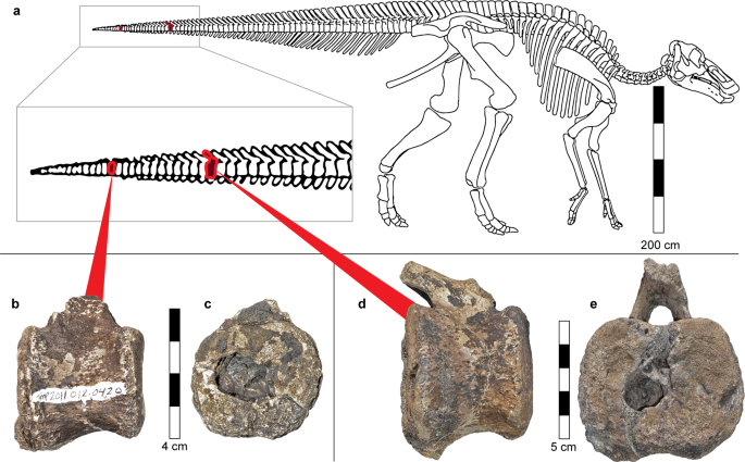
Rothschild, B., DePalma, R., Burnham, D. & Martin, L. Anatomy of a dinosaur – clarification of vertebrae in vertebrate anatomy. Anat. Histol. Embryol. In press, (2019).
El-Najjar, M. Y. Skeletal changes in tuberculosis: the Hamann-Todd Collection. Prehist. Tuberc. Am. 85–97 (1981).
Bierry, G. et al. Enchondromas in children: imaging appearance with pathological correlation. Skeletal Radiol. 41, 1223–1229 (2012).
Dabska, M. & Buraczewski, J. Aneurysmal bone cyst. Pathology, clinical course and radiologic appearances. Cancer 23, 371–389 (1969).
Dahlin, D. C. & Ivins, J. C. Fibrosarcoma of bone: a study of 114 cases. Cancer 23, 35–41 (1969).
Hudson, T. M. & Hawkins, I. F. Jr. Radiological evaluation of chondroblastoma. Radiology 139, 1–10 (1981).
Kransdorf, M. J., Moser, R. P. Jr. & Gilkey, F. W. Fibrous dysplasia. Radiographics 10, 519–537 (1990).
Milgram, J. W. The origins of osteochondromas and enchondromas. A histopathologic study. Clin. Orthop. Relat. Res. 264–284 (1983).
Shapiro, F. Ollier’s Disease. An assessment of angular deformity, shortening, and pathological fracture in twenty-one patients. J. Bone Joint Surg. Am. 64, 95–103 (1982).
Huvos, A. Bone Tumors: Diagnosis, Treatment, and Prognosis. (Saunders: Philadelphia, Pennsylvaniam USA, 1991).
Spjut, H., Dorfman, H., Fechner, R. & Ackerman, L. Tumors of Bone and Cartilage. (Armed Forces Institute of Pathology: Washington, DC, USA., 1983).
Bonakdarpour, A., Levy, W. M. & Aegerter, E. Primary and secondary aneurysmal bone cyst: a radiological study of 75 cases. Radiology 126, 75–83 (1978).
Rothschild, C., Rothschild, B. & Hershkovitz, I. Clues to recognition of kidney disease in archeologic record: characteristics and occurrence of leontiasis ossea. Reumatismo, 133–143 (2002).
Colmenero, J. D., Reguera, J. M., Fernandez-Nebro, A. & Cabrera-Franquelo, F. Osteoarticular complications of brucellosis. Ann. Rheum. Dis. 50, 23–26 (1991).
Rothschild, B. M., Hershkovitz, I. & Dutour, O. Clues potentially distinguishing lytic lesions of multiple myeloma from those of metastatic carcinoma. Am. J. Phys. Anthropol. Off. Publ. Am. Assoc. Phys. Anthropol. 105, 241–250 (1998).
Padgett, G. A., Madewell, B. R., Keller, E. T., Jodar, L. & Packard, M. Inheritance of histiocytosis in Bernese mountain dogs. J. Small Anim. Pract. 36, 93–98 (1995).
Reed, N., Begara-McGorum, I. M., Else, R. W. & Gunn-Moore, D. A. Unusual histiocytic disease in a Somali cat. J. Feline. Med. Surg. 8, 129–134 (2006).
Campione, N. E. & Evans, D. C. Cranial growth and variation in edmontosaurs (Dinosauria: Hadrosauridae): implications for latest Cretaceous megaherbivore diversity in North America. PLoS One 6, e25186 (2011).
Source: Ecology - nature.com


