Specimens examined
Twenty-three species of flies were assessed in this study. Specimens were obtained from the AgVic Victorian Agricultural Insect Collection (VAIC) or from recently trapped flies collected from northern Australia, through the Department of Agriculture, Water and the Environment (DAWE) Northern Australian Quarantine Strategy (NAQS) program. Species were initially identified morphologically using published literature1,2,17. The specimens examined, together with their known Australian distributions and pest status, are listed in Table 2. The species were chosen to include examples of the target species and the closest known relatives for testing the specificity of our novel LAMP assay, as well as a range of other Australian Tephritidae, Drosophilidae and Lonchaeidae pest and non-pest flies (Table 2).
DNA extractions
DNA was extracted from dry or ethanol (absolute) preserved adult whole specimens, larva, heads or legs of adults using DNeasy Blood and Tissue extraction kit (Qiagen), following the manufacturers protocol (Table 2). These samples provided “clean” DNA preparations to use for DNA barcoding for species identification and as positive controls for testing the LAMP assay. All specimens were identified morphologically, prior to DNA extraction. Species identification of all specimens (Table 2) was also confirmed using standard DNA barcoding methods4.
Additional “crude” DNA extractions were prepared from whole specimens of B. tryoni (eggs, larvae, pupae and adults) using QuickExtract DNA extraction solution 1.0 (Epicentre, USA). Fifty microliters of QuickExtract DNA extraction solution was pipetted into each 8 well strip of LAMP PCR tubes (OptiGene, UK). Fly samples were removed from ethanol and air dried on a paper towel for approx.1 min. Single whole adult fly (placed head down), larva, pupa, single and/or multiple eggs were pricked with an entomology pin (single use to prevent cross contamination), which was used both to help release DNA from specimens and transfer each specimen into a well. Each sample was immersed in QuickExtract DNA extraction solution with up to six samples processed simultaneously in the portable real-time fluorometer (Genie III, OptiGene, UK). The protocol used the Genie III as an incubator at 65 °C for 6 min followed by 98 °C for 2 min (Total run time = 8 min), following the QuickExtract manufacturers recommendations. The DNA was quantified using a NanoDrop ND-1000 Spectrophotometer (Thermo Fisher, Australia) and stored at −20 °C. Whole specimens post QuickExtract DNA extraction were retained and visually examined to confirm morphological identification features were still present.
Development and evaluation of LAMP assay
LAMP primer design
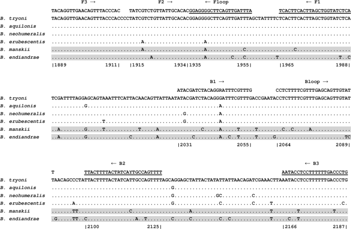
We developed new LAMP primers in this study to target the B. tryoni complex, including B. tryoni, B. neohumeralis and B. aquilonis. This assay was designed to target the 5′ region of the mitochondrial COI locus. All available sequences of the COI coding region of the target species were retrieved from the BOLD Systems public database and used for the LAMP primer design. A total of 54, 4 and 24 sequences were available for B. tryoni, B. neohumeralis and B. aquilonis respectively as of 11th September 2017. Alignments generated from these sequences are referred to as the “target” set. Reference fruit fly DNA sequences from the Bactrocera genus other than the target species of the B. tryoni complex were also retrieved from the BOLD Systems database (2922 sequences as of 11th September 2017) and used for comparison. The alignment generated from these non-target sequences is referred to as the “non-target” set. The sequences were aligned using the alignment program Clustal Omega provided at The European Bioinformatics Institute (EMBL-EBI; https://www.ebi.ac.uk/Tools/msa/clustalo/) using default parameters. Highly conserved regions within the target sequence alignments were identified manually, assessed for their suitability as LAMP primers and chosen based on the following criteria: i) sites with least degree of degeneracy within the target species yet still sufficiently different (preferably > 3 bases different) to the same region in the non-target sequence alignment; ii) sites with the least degree of self-annealing and loops formation, especially at the 3′ end and iii) their relative positions to each other in accordance with the design guidelines stated by the Primer Explorer software (http://primerexplorer.jp/e/). Primer degeneracy was added to account for known B. tryoni complex sequence variation based on the target alignment.
LAMP primer ratio optimisation
A primer master mix was prepared first for the assay for ease of reaction setup. The primer master mix was prepared by adding the specified amount of each of the six primers (Table 3) as per the ratio requirement for outer forward primer (F3), outer backward primer (B3), forward inner primer (FIP), backward inner primer (BIP), forward loop primer (Floop) and backward loop primer (Bloop). Due to the higher concentration required for the inner and loop primers, both were used at a concentration of 100 µM whereas the outer primers were diluted to a working concentration of 10 µM for ease of calculation. A 100 µL volume of primer master mix 1:6:3 (F3/B3: FIP/BIP: Floop/Bloop) was prepared by adding 10 µL each of F3/B3, 6 µL each of FIP/BIP, 3 µL each of Floop/Bloop and 62 µL of Ultrapure water (Invitrogen, Australia). The final concentrations of each LAMP primer within the newly developed assay were determined empirically based on published literature and previous work with LAMP (not published). The concentrations for the outer primers F3 and B3 were tested at 0.2 µM (5 µL of primer master mix and 5 µL of H2O), 0.3 µM (7.5 µL of primer master mix and 2.5 µL of H2O) and 0.4 µM (10 µL of primer master mix only) with the following ratios 1:6:3, 1:8:4, 1:10:5 and 1:12:6 for outer primers: inner primers: loop primers, for the B. tryoni LAMP assay. Optimisation of primer ratio is critical in the development of the LAMP assay, firstly to determine the optimum amplification time of target species (within 25 minutes), and secondly to be able to achieve a consistent annealing temperature for all the positive DNA samples. Negative samples and non-target species should not amplify and the anneal derivative should remain as a flat line.
LAMP assay conditions
Each LAMP reaction was conducted in a total volume of 25 µl containing 14 µl of Isothermal Master Mix (Iso-001, OptiGene, UK), 10 µl of the primer master mix containing all 6 primers at the appropriate concentrations and 1 µl of template DNA. A one microliter disposable plastic loop was used to add “crude” QuickExtract DNA, ensuring that a fresh loop was used for each sample. The use of disposable plastic loops eliminates the need for pipettes when LAMP is used outside a laboratory, a necessity for effective field deployment.
“Clean” DNA extracted from the target species using the DNeasy Blood and Tissue extraction kit was used as positive controls for the LAMP assay. A no template control (NTC) was included in each test to detect reagent contamination. All the LAMP assays were run in the Genie III at 65 °C for 25 min followed by an annealing curve analysis from 98 °C to 73 °C with ramping at 0.05 °C/s. The total run time is approximately 35 minutes.
Once the run has finished, the amplification and anneal derivative curves can be visualised on the Genie III screen to ensure that amplification has occurred as expected. Non-target species and NTCs are expected to have flat amplification lines. The time of amplification (minutes) and anneal derivative temperature (°C) are recorded from the Results Table displayed by the Genie III. The amplification time and the anneal derivative temperature of all the samples are compared against the positive control/s to confirm that no false-positive or false-negative has amplified.
The date, Genie III serial number and the run number of each LAMP assay completed on the machine was recorded so that the run files could be transferred and analysed using a PC version of the software Genie Explorer version V2.0.6.3. LAMP assays in this study were visualised in the blue channel of the Genie III.
Analytical sensitivity of the LAMP assay compared with qPCR
A five-fold serial dilution of a “clean” DNA extract was prepared using ultrapure water from two biological replicates of B. tryoni. Starting DNA concentrations were quantified using a NanoDrop ND-1000 Spectrophotometer (Thermo Fisher, Australia). B. tryoni DNA was serially diluted from 10 ng/µl to 0.016 ng/µl (1:1 to 1:625). Sensitivity of the LAMP assay was tested using the serially diluted DNA in the Genie III, following the same assay conditions as mentioned above. The time of amplification and anneal derivative temperatures were recorded for all samples.
The same serial dilution of DNA extracts was also used in real-time qPCR assay. The primers (manufactured by Sigma), probes (manufactured by Applied Biosystems and Sigma) and cycling conditions used were as published for Medfly8, including lowering the annealing temperature to 58 °C. Real-time qPCR was performed in a Rotor-Gene Q (Qiagen, Australia) in a total volume of 25 µl. Each reaction mixture included 12.5 µl Platinum Quantitative PCR SuperMix-UDG (Invitrogen, Australia), 0.5 µM of each forward and reverse primers, 0.2 µM Taqman probe, 5 µl of template DNA and made up to 25 µl with RNA-free water. An NTC with 5 µl of water instead of DNA was included in each run to check for reagent contamination. The thermal cycling conditions consisted of a two-step denaturation: 2 min at 50 °C and 10 min at 95 °C, followed by 35 cycles of amplification in a two-step procedure: 95 °C for 10 seconds and 58 °C for 1 min. The Cq value (cycling quantification value) of the 5 dilutions for the two biological replicates of the target species were recorded for comparison with the time of amplification obtained from the LAMP assays.
Source: Ecology - nature.com


