Systematics
Thylacocephala sensu 21
Concavicarida sensu10
Concavicaris sensu22.
Diagnosis
“Optic notches generally modest in size, prominently defined, dorsally delineated by a variously developed rostrum, ventrally by variously developed anteroventral processes. Posterior aspect of carapace truncated but gently rounded” (Schram, 201423).
Remarks
C. submarinus is assigned to the genus Concavicaris based on the following morphological characters: optic notch modest in size and the posterior of the carapace is truncated and gently rounded. Additionally, the rostum and anteroventral process are variously developed among the genus, which does not disagree with the rostrum and anteroventral process observed in C. submarinus.
Species Concavicaris submarinus sp. nov.
Etymology
From the Latin word submarinus referring to submarine, as their shape is reminiscent of these watercrafts.
Holotype
PIMUZ 37349, which is an exceptional specimen preserving both eyes, posterior limbs and some internal structures (visible in the CT-images).
Paratypes
PIMUZ 37348, PIMUZ 37350, PIMUZ 37351, PIMUZ 37353, PIMUZ37354, AA.MEM.DS.3, AA.MEM.DS.4 and AA.MEM.DS.5.
Nomenclatural statement
A Life Science Identifier (LSID) was obtained for the new species (C. submarinus): urn:lsid:zoobank.org:act:XX, and for this publication: urn:lsid:zoobank.org:pub:XX.
Material
Although there are many fossils in the field and some tens of specimens were collected, we selected the best preserved specimens for the description (PIMUZ 37348–51, PIMUZ 37353–4 and AA.MEM.DS.3, AA.MEM.DS.4 and AA.MEM.DS.5). The size of the specimens varies from 27 mm (PIMUZ 37354) to 98 mm (PIMUZ 37348) in length and 10 mm (PIMUZ 37354) to 46 mm (PIMUZ 37348) in height.
Locality and horizon
Madène El Mrakib, Morocco; Maeneceras genozone, middle Famennian, Late Devonian.
Diagnosis
The optic notch has a small anteroventral process. The mid-ventral margin of the carapace curves towards the outside. A depression is visible along the curved margin, beginning shortly after the curve anteriorly and forming a longitudinal line until it reaches about 2/3 of the carapace length. There is no sign of ornamentation except the polygonal pattern of the cuticle.
Description
The holotype, PIMUZ 37349, is 56 mm long, 26 mm high and 15 mm wide (Fig. 1a). The biggest specimen available to us, PIMUZ 37348, lacks the posterior end, but its total length can be estimated to have reached nearly 100 mm at a height of 46 mm (Fig. 1c). PIMUZ 37354 lacks the rostrum and is the smallest specimen, which is 27 mm long and 10 mm high (Fig. 1f). AA.MEM.DS.4 and PIMUZ 37354 lack their rostrum. All specimens reach the maximum carapace height anterior to the centre.
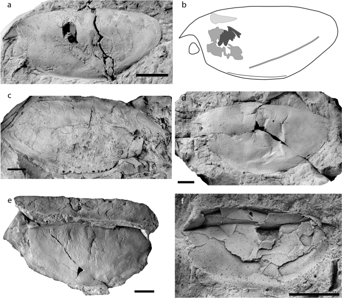
Specimens of Concavicaris submarinus. (a) Holotype PIMUZ 37349. (b) Line drawing of the holotype, showing the location of the ventral curve, the depression lying from anterior the rear end until midway through the carapace, and the internal structures (section of the gills in black, putative gastric muscles in dark grey and paired structure in light grey). (c) PIMUZ 37348, the biggest specimen, lacks the rear end. (d) PIMUZ 37350. (e) PIMUZ 37353. (f) PIMUZ 37354, the smallest specimen. Anterior is on the left for (a,b,d,f). Anterior is on the right for (c,e). Scale bars indicate 10 mm.
There is no posterior spine in any of the specimens. When the rear end is preserved, it appears always truncated and gently rounded. The anteroventral process of the optic notch is small in size and visible in most specimens (Fig. 1).
The most noticeable diagnostic feature of Concavicaris submarinus lies on the ventral margin (Figs. 1 and 2). This edge has a section anterior to the centre, which is bent outwards, i.e. laterally (e.g. Fig. 1a,d,e). This is best visible in the least deformed specimens (AA.MEM.DS.3 and 4, PIMUZ 37348, 37349, 37353, 37354). Along the ventral margin, a depression runs longitudinally from the anterior starting point of the bent margin along a third of the carapace length (Fig. 1a,b).
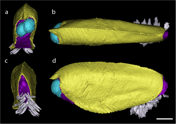
3D reconstruction of PIMUZ 37349, the holotype of Concavicaris submarinus. (a) frontal view. (b) posterior view. (c) dorsal view. (d) lateral view. The carapace is here in yellow, the main body appears in purple, the eyes are in blue and the paddle-like limbs in white. Scale bar indicates 10 mm.
The large compound eyes of Concavicaris submarinus are well preserved in the holotype and reach a seventh of the body length (see on Fig. 2). Their structure is clearly visible under the microscope and shows a clear and regular pattern of convex, slightly elongated, hexagonal facets (Fig. 3a–c).
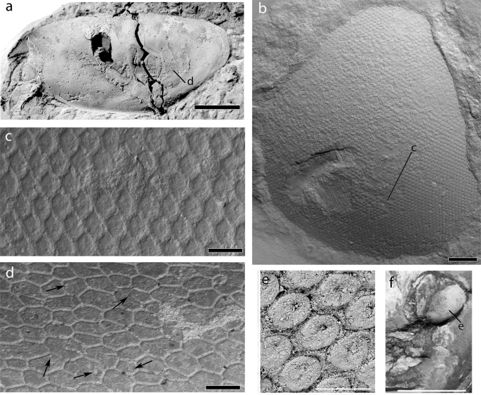
SEM images of the eye surface structure and cuticle of the carapace of the holotype of Concavicaris submarinus n. sp. (PIMUZ 37349; a–d) (a) the entire specimen, coated with NH4Cl-sublimate. (b) detail showing of the eye. (c) close-up of the hexagonal facets of the eye. (d) a section of the cuticle, visible on most of the carapace, made of polygons, some of which show a round depression (indicated by black arrows). (e) close up of the hexagonal facets of the eye of Dollocaris. (f) detail showing of the eye of Dollocaris and place where (e) was made. (e,f) were modified from26 Scale bars are (b) 500 µm, (c) 100 µm, (d) 200 µm, 50 µm (e) and 10 mm (f).
The carapaces of eight specimens preserve the cuticle in several places. Its good preservation in various areas of the carapace (e.g., PIMUZ 37349) reveals the polygonal imprints of cuticle cells, bordered by thin grooves (Fig. 3d). The polygons are irregular in the number of sides, overall shape and size. Some cells also show a more or less central depression (see black arrows on Fig. 3d).
In PIMUZ 37349 eight pairs of paddle-like limbs are preserved at the posteroventral end (only visible in the CT-images) – one leg per side being incomplete (Fig. 2). The legs on its left side are taphonomically deformed, making them appear much thicker (6.1 mm compared to 2.4 mm on the other side) than long (6.5 instead of 14 mm on the other side). Because of the deformation, it is conceivable that the legs have been pushed outwards, which could explain why many of them seem to be detached from the body. The legs expand from posterior of the curved ventral margin until shortly posterior of the carapace where they reach out of the carapace. The trunk segments are not visible in the scan (Fig. 4), either because they did not preserve or because of an insufficient density contrast.
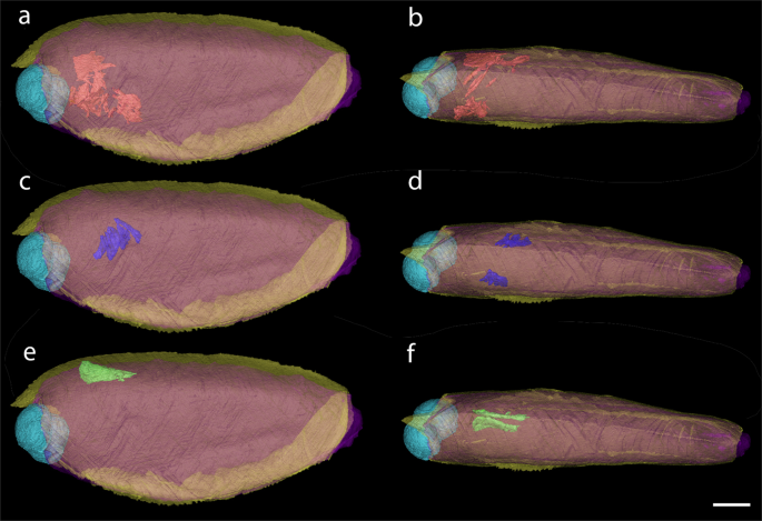
Internal structures of Concavicaris submarinus (PIMUZ 37349). The presumed gastric muscles appear here in red and are seen in a lateral (a) and dorsal (b) view. The sections of the putative gills are shown in blue, in both lateral (c) and dorsal (d) view. The elongated structures, interpreted as gonads or the hepatopancreas are shown in green and in a lateral (e) and dorsal (f) view. Scale bar is 10 mm.
One of the most remarkable features seen in the holotype is the presence of thin structures on both anterior sides of the specimen (Fig. 4a,b). They appear to surround the area where the stomach is assumed to be, which could imply that these thin structures could be gastric muscles.
The CT scan revealed another structure near the putative gastric muscles. Three pairs of lamellar structures are visible (Fig. 4c,d). They are thin, divided in leaf-like sections and resemble gills.
Additionally, a pair of elongated organs are located dorsally to the presumed gastric muscles and anteriorly to the supposed gills (Fig. 4e,f). There is a clear separation between the two parts of this tissue, thus suggesting that it is a paired structure.
Remarks
Thylacocephalans are undoubtedly arthropods, but their affinities within this clade are still widely debated1,2,3. This lack of systematic resolution roots in the very incomplete knowledge of thylacocephalan anatomy. Therefore, exceptionally preserved materials such as those presented here can provide valuable anatomical details helping to better resolve trees comprising thylacocephalans. In the following, we discuss the structures that became visible in the CT-scan.
The cuticle displays a polygonal cuticular pattern separated by thin grooves, which are characteristic of thylacocephalans9. Secrétan24 and Broda et al.9,25 also suggested that the presence of a central depression in these polygons, the lumen (Fig. 3d), may represent pores where sensory setae inserted.
The eyes of C. submarinus have features strongly resembling the eyes of the Jurassic thylacocephalan Dollocaris ingens from La Voulte26, although the eyes of Dollocaris are significantly larger. Similarly, the Moroccan material has eyes composed of a dense and relatively regular pattern of hexagonal facets (Fig. 3b,c). However C. submarinus shows facets a little bit more elongate and a more hexagonal shape than Dollocaris in a similar surface area of the eye (Fig. 3e,f). The facets are also convex while those of Dollocaris appear to be more concave, probably due to post-mortem collapse26.
The thin and flattened, yet slightly inward curved structures (Fig. 4a,b) resemble muscles. The location on these structures (anterior and lateral) implies that there could have been an organ or other internal structure between. The position of the putative muscles surrounds the space where we would expect the stomach3,26. Thus, we interpret these structures as gastric muscles. Further remains of musculature are found on the anterior right side of the holotype. They lie above the area where the stomach likely used to be. We think that this could be remains of an anterior gastric muscle. Among the presumed gastric muscles (Fig. 4a,b), some can be tentatively interpreted as external mandible adductor muscles, similar to what has been described from decapod crustaceans27, but the possibility that these could be lateral gastric muscles cannot be ruled out until better preserved specimens reveal more anatomical details. No mandibles or other mouth parts have been reported from thylacocephalans so far, implying that the gastric muscle interpretation is more probable than being part of the mouth parts.
The leaf-like lamellar structures show three branches on each side (Fig. 4c,d). This could imply paired organs that are close to one another. The position of these structures is compatible with their interpretation as gills. However, gills normally should consist of eight pairs of lamellae, which is characteristic of thylacocephalans. Also, they are not as long as expected. This suggests that some lamellae are probably simply not preserved, just like most other internal organs and structures, or they were not visible in the CT-imagery due to an insufficient contrast in density.
The shape of the paired structure segmented in green (Fig. 4e,f) resembles that of gonads in some decapod crustaceans, which also concurs with the phylogenetic position of thylacocephalans. Kienbaum et al.28 discussed the gonads of decapods and explained that the ovaries are a pair of elongated organs located dorsally in the cephalothorax, a description fitting well with the structures found in the holotype. Although it is expected that gonads appear further posteriorly, a few species seem to have them more anteriorly than thought. Nagaraju29 detailed the same physiology and position in the body as Kienbaum et al.28, and located the ovaries on top of the stomach and hepatopancreas in some decapod crustaceans. The description of the hepatopancreas in crustaceans30 could fit the location of such structure, but this can be omitted here because thylacocephalans have a large hepatopancreas located ventrally in the thorax26. Although the gonad hypothesis stands a bit more than other interpretations like the hepatopancreas, further findings will contribute to revealing the correct interpretation of this structure.
C. submarinus differs from all concavicarids with its near mid-ventral fold. Species like C. milesi has a deep “U”-shaped optic notch10, which is not the case in C. submarinus. Also, the cuticle of C. milesi is terraced, unlike that of C. submarinus. C. sinuata has a deep carapace and a thick rostrum that extends to the anteroventral process10. This is not visible in C. submarinus. C. submarinus has a shorter carapace than C. elytroides10,31 and both have different types of cuticle; the cuticle of C. elytroides is striated while that of C. submarinus is much more similar to C. bradleyi. C. submarinus is a close relative of C. bradleyi31. They both have a convex carapace with a broad arch forming the dorsal margin. Both species have a similar outline with a rounded posterior end, a minute anteroventral process and a moderately-sized rostrum. Three differences distinguish these two species. First, the ventral margin of C. submarinus curves outwards while in C. bradleyi, the subcentral ventral margin is inflected inwards and upwards31. Secondly, C. submarinus shows a depression on the carapace reaching from near the curve to almost the rear end. Thirdly, the interior angle of the eye is sharper in C. submarinus than in C. bradleyi, here it is more rounded.
Source: Ecology - nature.com
