Description of the study area and plant collection
Entire flowers were manually collected from endemic plants growing on the banks of the Nema River, next to Dielmo (13°43′23.43′′N, 16°24′46.27′′W) and Simong (13°37′55.63′′N, 16°23′08.24′′W) (Fig. 3). Dielmo is located in the Fatick region of Senegal, in an area of Sudanese savannah approximately 174 miles from Dakar and 9.32 miles north of the Gambian Republic. Since 1990, the area near Dielmo has hosted one of the oldest cohort studies investigating the epidemiological relationships between the malaria parasite and its vectors and human hosts50. Previous studies have already laid out the geographical and epidemiological characteristics as well as the dynamics of malaria in Dielmo50,51.
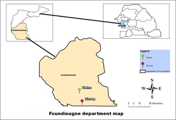
Map of the study sites. The positions of the two collection sites are indicated by red (Simong) and green (Dielmo) dots. Both villages are close to the Saloum delta, one of the most irrigated areas in Senegal.
Members of the An. gambiae s.l. and An. funestus group are the main vectors of malaria in this area52,53.
The flowers were collected from 27 to 30 October 2016. They were cut at the stalk level, placed in bags (one bag per plant), stored at 4 °C in the Dielmo laboratory and transported to Dakar at the same temperature. In Dakar, the samples were immediately frozen at −80 °C until they were processed to assess their molecular biology and for bacterial cultures.
Molecular analyses
DNA extraction
The petals and sepals of each flower were removed, and then the peduncle was cut off. The remaining flower parts, including nectar and pistil, were immersed in an Eppendorf tube containing 600 μl of 1X phosphate buffered saline (PBS) and then crushed. After centrifugation, 200 μl of the supernatant was collected for DNA extraction, which was performed according to the 2% CTAB method54 supplemented with β-mercaptoethanol at a concentration of 0.2%. The mixture was left to digest overnight. The same DNA extraction procedure was performed for the mosquitoes. Before DNA extraction, individual specimens were sacrificed at −80 °C, washed with 70% ethanol to remove superficial environmental bacteria and then crushed. Then, genomic DNA was immediately extracted from the whole body of each individual specimen as previously described54.
Asaia bogorensis molecular detection
Taqman® qPCR methods with custom designed primers and probe sets were used to screen samples for the presence of Asaia spp. by targeting the rpoB gene (Fwd: 5′-GACGCCAAGGACCTGATCTA-3′; Rev: 5′-ATAGGCCAGGATTTCGTCCT-3′; Probe: 6-FAM-GGTCACGACCCTGCTCTATG-TAMRA)2. The targeted region was amplified in a total reaction volume of 20 μl, containing 10 μl of ROCHE® master mix (Roche Diagnostics, Indianapolis, IN, USA), 0.5 μl of each primer, 0.5 μl of the probe, 3 μl of distilled water, 0.5 µl of UDG, and 5.0 μl of test DNA. The conditions of the amplification with the CFX96 Touch detection system (Bio-Rad, Marnes-la-Coquette, France) were as follows: 2 minutes at 50 °C for UDG action followed by an initial denaturation of 5 minutes at 95 °C, then 39 cycles of 5 seconds at 95 °C and 30 seconds at 60 °C. A sample was considered positive when the Ct value was less than 35 cycles. The Anopheles negative control was prepared from a crush of certified Asaia-free An. coluzzii, and the positive control was prepared from An. coluzzii to which we added a suspension of A. aff. bogorensis GD01.
16 S rRNA gene sequencing and phylogenetic tree construction
The molecular phylogenetic evolutionary history was inferred from a nearly complete 16 S rDNA sequence (1249 bp), which was sequenced using universal 16 S rDNA primers fD1 and rp255, as described by Drancourt et al.56.
Initially, we used a Peltier PTC-200 model thermal cycler (MJ Research Inc., Watertown, MA, USA) to perform the PCR amplifications. The reactions were carried out using HotStar Taq polymerase (Qiagen, Hilden, Germany) following the manufacturer’s instructions. For each assay, we included negative and positive controls for the validation of the run. The success of amplification was confirmed by electrophoresis on a 1.5% agarose gel.
The purification of the PCR products was performed using NucleoFast 96 PCR plates (Macherey-Nagel EURL, Hoerdt, France) according to the manufacturer’s instructions. The products of the amplification were then sequenced using a Big Dye Terminator Cycle Sequencing Kit (Perkin Elmer Applied Biosystems, Foster City, CA) with an ABI automated sequencer (Applied Biosystems).
The amplified sequences were assembled and corrected using ChromasPro software (ChromasPro 1.7, Technelysium Pty Ltd., Tewantin, Australia) and then compared with reference sequences available in Genbank using the BLASTN server (http://blast.ncbi.nlm.nih.gov/Blast.cgi) to identify closely related species and/or strains.
The taxonomic relationships of the new strains were inferred against the existing isolates. The reference sequences retrieved from the Genbank database, together with the Senegalese strains, were aligned using the ClustalW multisequence alignment program57 in BioEdit software58. A maximum likelihood phylogenetic tree was reconstructed using TOPALi v2.5 based on the Hasegawa-Kishino-Yano (HKY85) substitution model59, which includes the proportion of invariable sites and the gamma distribution. The robustness of the individual branches was estimated by bootstrapping with 100 replicates60.
Establishment of an Asaia-free An. coluzzii strain
Sensitivity of A. aff. bogorensis GD01 to antibiotics
The sensitivity of the A. aff. Bogorensis GD01 strain, previously isolated from An. gambiae from Dielmo village2, to antibiotics was determined. A pure suspension of A. aff. bogorensis GD01 at a concentration equal to the 0.5 McFarland standard was inoculated onto Mueller-Hinton agar medium61,62. Antibiotic (amoxicillin, amoxicillin/clavulanic acid, piperacillin/tazobactam, doripenem, imipenem, aztreonam, ceftriaxone, ceftazidime, cefpirome, gentamicin, doxycycline, erythromycin, trimethoprim/sulfamethoxazole, ciprofloxacin, linezolid, metronidazole and rifampicin) discs were deposited onto dried agar plates and the plates were then incubated at 28 °C under 5% CO2 atmosphere. Finally, the inhibition diameters were measured and compared with the reading charts according to the manufacturer’s instructions (i2a – Siège Social, 401 Avenue du Walhalla, CS83406, 34060 Montpellier Cedex 2,France) and the recommendations of the Antibiogram Committee of the French Society of Microbiology63.
Isolation of strains of Asaia spp. from wild plants
Suspensions of the qPCR-positive I. pes-caprae were seeded on Columbia agar supplemented with 5% fresh sheep blood and 4 mg/l gentamicin (batch No. F140512, manufactured by Xin K. Pharm Co. Ltd). The seeded medium was then incubated at 28 °C under an atmosphere of 5% CO2. The colonies morphologically resembling Asaia were seeded on another Columbia agar plate supplemented with 5% fresh sheep blood but without antibiotics and incubated under the same conditions. Approximately 50 purified colonies were then collected and suspended in 200 μl of sterile 1X PBS and used for bacterial DNA extraction using the 2% CTAB method supplemented with 25 μl of proteinase K (Tritirachium album, 25 mg, ref. EUC0090-A, EUROMEDEX, 24, rue des Tuileries BP684 67460 Souffelweyersheim, France). Finally, the extracted DNA was screened for the presence of Asaia using A. bogorensis-specific qPCR.
Treatment of adult mosquitoes with antibiotics
Adult An. coluzzii colonies maintained at a temperature of 26 ± 2 °C and a relative humidity of 80 ± 10% in our laboratory, as described previously50,51,52,53, were fed a mixture of sterile water containing doxycycline (batch No. B113304 manufactured by SERB Laboratories) at a concentration of 8 mg/l and 10% sucrose solution for 4 days. qPCR was performed on the treated mosquitoes to confirm their Asaia-free status.
Insectary rearing of the Asaia-free mosquito strain
The Asaia-free mosquito line, fed sugar solution prepared with autoclaved water, was maintained at a temperature of 26 ± 2 °C and a relative humidity of 80 ± 10%. When feeding the mosquitoes, a volume of 50 ml of autoclaved water was filtered with a 0.2 µm filter and then mixed with sterile sucrose solution at a concentration of 10%. This mixture was offered daily to mosquitoes and changed daily to prevent mosquito reinfection. The physical conditions of breeding were the same as those of the other mosquitoes in the insectarium. The control of the absence of Asaia in this mosquito line was assessed weekly and before each experiment using our Asaia-specific custom-designed qPCR system.
Design of the experimental infection through transmission of A.aff. bogorensis
The two An. coluzzii colonies (Asaia-infected and Asaia-free), maintained separately in the insectarium of the VITROME laboratory of the Institut de Recherche pour le Développement (Dakar, Senegal), were used for the experimental infection of A. aff. bogorensis transmission. The common ornamental plant P. reticulatum (Acanthaceae) was selected as the source of nectar during the experimental infection study after the confirmation of its natural Asaia-free status and due to its easy procurement from urban horticulturists in Dakar, where our laboratory is based. The plants were grown in pots within a closed room with no access to flying arthropods to prevent the contamination of newly opened flowers with Asaia. Randomly selected flowers from each plant were screened for the presence of Asaia spp. and only plants that were negative were used for the subsequent experimental infection.
Overall, 310 mosquitoes of the naturally Asaia-infected laboratory colonies (123 males and 187 females) were brought into contact with an uninfected P. reticulatum bearing newly opened flowers. The plant in its pot was introduced into a closed 100 × 50 × 50 cm mosquito cages for 3 days. During the experimental infection, the average temperature of the standard thermo-hygronomic device was 26 ± 2 °C, and the relative humidity was 80 ± 10%. At the end of the third day, the mosquitoes were sacrificed at −80 °C and then screened with randomly selected flowers using our qPCR method to confirm that the mosquitoes and the flowers of the exposed plants were infected with Asaia. The following day, an unfed batch of mosquitoes from the Asaia-free lines was allowed to feed for 3 days on the Asaia-infected plants under the same conditions described above. After exposure, the mosquitoes were sacrificed, stored individually in Eppendorf tubes for DNA extraction and then examined for Asaia infection. This experiment was repeated twice with the same number of anopheles for both lineages and the same plant species under identical physical conditions.
Statistical analyses
This study was carried out over two consecutive years. Two groups of mosquioes were used each time. This first group was the An. coluzzii strain naturally infected by A. aff. bogorensis and the second was as An. coluzzii Asaia-free group obtained after antibiotic treatment and then exposed to infected P. reticulatum. Within each group, we analyzed the differences in infection rates between the two sexes. Statistical analyses were performed with Epi Info software version 7.0.8.8.0 (Centers for Disease Control and Prevention, Atlanta, GA, USA). The differences were analyzed using the Yates corrected χ2 test with one degree of freedom and a 95% confidence interval. The difference was considered significant when the bilateral p value < 0.05
Source: Ecology - nature.com
