It has previously been shown in acute 24 h tests that small (50 to 60 nm) positively charged aminated polystyrene nanoparticles (PS-NH2) are the most toxic particles among the polystyrene nanoparticles tested on D. magna24. Therefore, 53 nm PS-NH2 nanoparticles were chosen in the present study to determine the lowest concentrations of nanoparticles observed causing mortality of D. magna in life-time exposure. Two to five day-old D. magna were isolated and exposed to polystyrene nanoparticles (Fig. 1) throughout their entire life-time, which for the oldest animal was 103 days. A concentration of 0.32 mg/L was chosen based from preliminary studies with aminated polystyrene nanoparticles (data not shown). In order to compare differently charged nanoparticles of specific surface areas, we increased concentrations for 62 and 26 nm carboxylic modified particles.
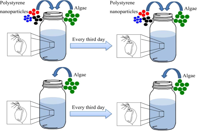
Schematic representation of long-term toxicity test. In total, there were ten replicates in each treatment. During the exposure to polystyrene nanoparticles, alive Daphnia magna individuals were transferred every third day to 100 mL glass beakers with 80 mL total volume of fresh medium, containing 2.5 mL of food (algae), with (treatment) or without (control) particles. Nanoparticles were dialyzed prior the experiments and particle sizes were measured during exposure using DLS. Algae concentration and water pH values were measured every time D. magna was transferred.
D. magna individuals exposed to 0.32 mg/L of 53 nm PS-NH2 showed an increased mortality (χ2(1) = 10.19, p < 0.01) compared to the control group, while lower concentrations (0.032 and 0.0032 mg/L, Fig. 2) did not have any significant effects (χ2(1) = 0.89 and 0.089, respectively, p > 0.05, Fig. 2). The lowest lethal concentration in the present study (0.32 mg/L) was 78 times lower compared to the lowest lethal concentration (25 mg/L) previously used in acute tests24.
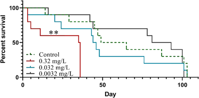
Survival of Daphnia magna exposed to different concentrations of 53 nm PS-NH2 throughout their life-time. Asterisk indicates significant difference compared to the control group estimated over the whole study period, **p < 0.01.
We also addressed the question if polystyrene nanoparticles that did not induce mortality in acute test24 is toxic at long-term (life-time) exposure. Negatively charged carboxylated polystyrene nanoparticles (PS-COOH) at the sizes 26 and 62 nm have been shown to be non-toxic in 24 h acute tests at concentrations up to 400 mg/L24. However, after long term exposure to lower concentrations (7.6, 3.2, 0.76 and 0.32 mg/L) of 62 nm PS-COOH in our study, D. magna showed a significant decrease in survival (χ2(1) = 3.85, 8.03, 4.55 and 6.89, respectively, p < 0.05, Fig. 3a). Similarly, D. magna showed a significantly reduced survival rate than in the control when exposed to both 3.2 and 0.32 mg/L of 26 nm PS-COOH (χ2(1) = 4.51 and 5.04, respectively, p < 0.05, Fig. 3b). For none of the sizes sub-lethal concentrations were reached and we may therefore conclude that although these carboxylated polystyrene particles were not considered toxic at short-term 24 h exposure24, they are indeed lethal at similar concentrations as the aminated particles at prolonged exposure.
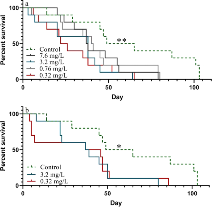
Survival of Daphnia magna exposed to 62 nm PS-COOH (a) and 26 nm PS-COOH (b) particles throughout their life-time. Asterisks indicate significant differences throughout the study period compared to the control group, *p < 0.05, **p < 0.01. Asterisks added on the control group indicate that all treatments were significantly different from the control group.
Interestingly, there was an apparent reversed concentration dependency in survival between the lowest (0.32 mg/L) and highest (7.6 mg/L) concentrations of 62 nm PS-COOH. It could be speculated, especially as the polystyrene nanoparticles are mixed with the algae, that the exposure scenario was influenced by differences in nanoparticle concentrations, e.g. aggregation and/or faster sedimentation. Sedimentation was shown to be an important factor affecting exposure scenarios in a life-time test evaluating the effects of tungsten carbide nanoparticles38. However, in the present study, no sedimentation was observed over 48 h at a particle concentration of 7.6 mg/L mixed together with algae (Fig. S1). Furthermore, no particle aggregation, measured by dynamic light scattering (DLS), was observed in neither the lowest, nor the highest concentrations (Table S1). Another possible difference in exposure scenario is that the binding of organic molecules to the particle surfaces changes the toxicity of the particles. This effect has been shown for polystyrene particles pre-incubated in algae and in media containing molecules secreted from D. magna29,33. Increasing the particle concentration from 0.32 to 7.6 mg/L causes an increase in added particle surface area from 2.4 × 10¹¹ to 56.4 × 1011 µm2 which may affect which type and how much organic material is bound to the particles.
A comparison between the effect of 53 nm PS-NH2, 62 nm PS-COOH and 26 nm PS-COOH at 0.32 mg/L, revealed a significant difference in the survival of D. magna between 53 nm -NH2 and 26 nm -COOH treatments (χ2(1) = 3.88, p < 0.05, Fig. 4). This implies that although the PS-COOH was shown to be toxic in the life-time experiments, but not in acute tests24, there is still a charge dependent toxicity. Generally, the positively charged PS-NH2 have been shown to be more toxic to D. magna, which might be due to a stronger interaction with the negatively charged Daphnia cell membrane29. It has also been shown that 50 nm PS-NH2 particle induces apoptosis in a variety of cells, while negatively charged nanoplastic particles did not have a significant effect39.
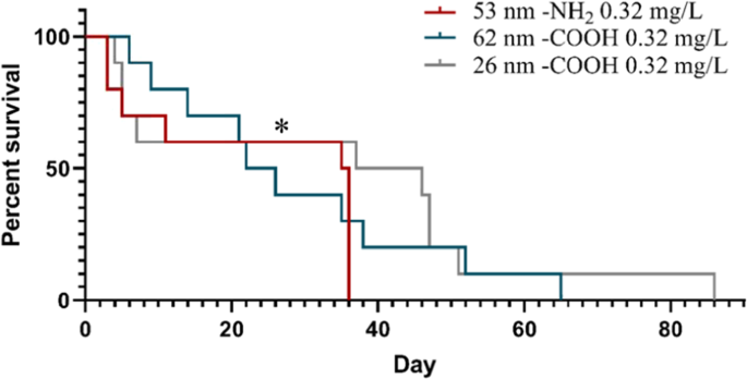
Comparison of Daphnia magna survival during life-time exposure to 53 nm PS-NH2, 62 nm PS-COOH and 26 nm PS-COOH at 0.32 mg/L. Asterisk indicates significant difference compared to the control group throughout the study period, *p < 0.05.
The accumulation of polystyrene nanoparticles in the body of D. magna has previously been demonstrated using fluorescent nanoparticles29, including the uptake of 20 and 70 nm particles25,40, as well as the accumulated body burden after 21 days exposure to 100 nm fluorescent polystyrene particles37. However, no data is available for the accumulation of non-fluorescent polystyrene nanoparticles. In order to document any microscopic changes during the life-time exposure, microscopic images were taken after death of several randomly chosen D. magna individuals (n = 3 for each treatment) that died after 30 to 100 days of exposure to different concentrations and sizes of PS-NH2 and PS-COOH. In some of the D. magna exposed to 53 nm PS-NH2 and 62 nm PS-COOH the gut contents were blackish (Fig. S2-B-C), which was not seen in individuals exposed to 26 nm PS-COOH (Fig. S2. D). These observations might suggest an accumulation of nanoplastic particles in some of the exposed organisms. This was not observed in any of the photographed individuals from the control group, where the guts instead had greenish contents from algal feeding (Fig. S2. A). Accumulation of nanoplastics in the gut of several organisms has previously been observed. For example, Torre et al.41 noted that after 48 h exposure negatively charged particles were accumulated in the digestive tract of sea urchin embryos, whereas positively charged nanoplastic particles were more dispersed in the gut. Nanoplastic particles aggregates have also been observed in D. galeata exposed to 52 nm polystyrene nanoparticles35. Similarly, Jemec et al.28 showed that polyethylene terephthalate textile microfibers were present in the gut of tested D. magna after 48 h exposure. Microplastic particles were also seen in the gut of exposed D. magna after 24 h test to particle concentrations of 12.5–400 mg/L, while the guts of control animals were greenish34.
In our study, the total number of offspring produced during the whole exposure time in treatments and the control group were not significantly different, neither by nanoparticle size nor concentration used within the same time period (p > 0.05 one-way ANOVA, Table S2). Similary, Rist et al.37 showed that reproduction was not effected after 21-day exposure to micro- and nanoplastic particles. However, at increasing concentrations of polystyrene nanoparticles, there was a decreasing trend in the number of offspring over their life-time (Table S3). Similarly, Besseling et al.33 also observed that increasing concentrations reduced the number of D. magna offspring. D. magna exposed to 0.1 mg/L of 1–5 μm microplastics of polymer microspheres for 21-day showed a significant reduction in reproduction42. Rist et al.37 showed that there was no difference in time to first offspring when D. magna were exposed to micro- and nanoplastic particles for 21 days, whereas Pacheco et al.43 observed a delay in the first brood release in D. magna after exposure to 1–5 μm microplastics. Likewise, Ekvall et al.38 showed a significant delay in time to first brood in D. magna exposed to tungsten carbide nanoparticles.
The majority of the published studies focus on acute, short-term, tests at high plastic particle concentrations24,29,34, whereas long-term toxicity studies on nanoplastics are rare, despite long-term, even life-time, exposure to low concentrations is the rule as nanoparticles enter natural ecosystems. Therefore, our understanding on how life-time exposure to nanoplastic particles affect organisms in aquatic food chains still remains elusive. Potential effects on aquatic organisms, such as zooplankton, may have considerable consequences for the function of aquatic food webs in which these organisms play a key role. In natural environments aquatic organisms are exposed to different sizes of plastic particles during their whole life-span. Our life-time experimental set-up does not only demonstrate toxicity of nanoplastic particles at relatively low concentrations, but also reveals toxicity of nanoplastics that are apparently non-toxic in standardized 24 or 48 h acute tests even at very high concentrations. Furthermore, in many cases mortality occurs after the standardized long-term 21-day tests. This clearly suggests that routine, standard test times may not be enough to assess the severity of plastic particles in our environment. Hence, by introducing life-time exposure tests we were here able to identify lethal effects at concentrations almost two magnitudes lower than previously shown29. Moreover, mortality may not be optimal to assess the lowest concentration of nanoplastic particles that will negatively affect the environment. Slow uptake of nanoplastics at low concentrations allow for accumulation of particles by the individuals, whereas high concentrations of nanoplastics in acute tests may rip off tissue or deplete the digestive system of neseccary enzymes44,45,46. In the future there is a need for mechanistic studies of the long-term toxicity in order to be able to properly assess the environmental risk, as well as the risk of different kinds of plastic particles.
Although the relevant concentrations of nanoplastic particles have, due to methodological constraints, not been determined, we here use relatively low concentrations of nanoplastic. We conclude that long-term exposure to low concentrations of nanoplastics material may provide considerably different outcomes with respect to toxicity than short-term, acute tests at high concentrations. Since long-term, or even life-time exposures may even already be ongoing in many regions of the world, our results have considerable implications for our use and manufacturing of plastic materials.
Source: Ecology - nature.com