Diagnosis of Stenhelmoides Grouvelle, 1908 mature larva
Body elongate, subcylindrical, parallel sided; color yellowish brown to reddish brown; tegument surface with several spatulate and scale-like setae as long as antennae (excluding sensorium), especially on head, sternal region of thorax, legs and first abdominal sternite (Figs. 1–5). Head exposed or partially concealed by pronotum, capable of being retracted into prothorax; frontal lines Y-shaped, merging basally into a long coronal line, about 1/4 of head; stemmata absent; clypeus concave without anterolateral tooth; labrum oval, wider than long; antenna with three antennomeres, second antennomere longer than other two together, with antennal sensorium long, as long as or slightly longer than second antennomere; mandibles symmetrical, with three apical teeth; prostheca long; maxilla with stipes longer than palpus, with four palpomeres; galea entire; lacinia well-developed, fused to stipes; labium with postmentum three times longer than prementum, palpi with two palpomeres, inserted on a short palpiger. Prothorax with six ventral sclerites, one antero-lateral pair and two lateral pairs, procoxal cavities open and membranous; meso- and metathorax with seven ventral sclerites, three large anterior process-like sclerites and two lateral smaller pairs, coxal cavities open; meso- and metathoracic sternal sclerite strong and prominent between coxae. Abdomen with a single pair of pleural sclerites on first segment; sternopleural and tergopleural sutures incomplete on second abdominal segment; abdominal segments wider than long; segment IX ending in two spinous processes with an emargination in the middle. Ventral operculum on abdominal segment IX pentagonal. Abdominal hooks lacking teeth on inner margin.
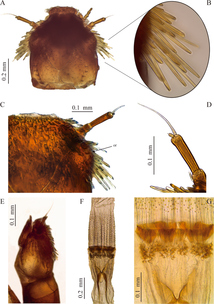
Head and mouthparts of S. rufulus larva. (A–C) Head, (A) dorsal view, (B) lateral spatulate setae, (C) frons and ocular region, (D) antenna, (E) head, lateral view, (F,G) proventriculus. or: ocular region.
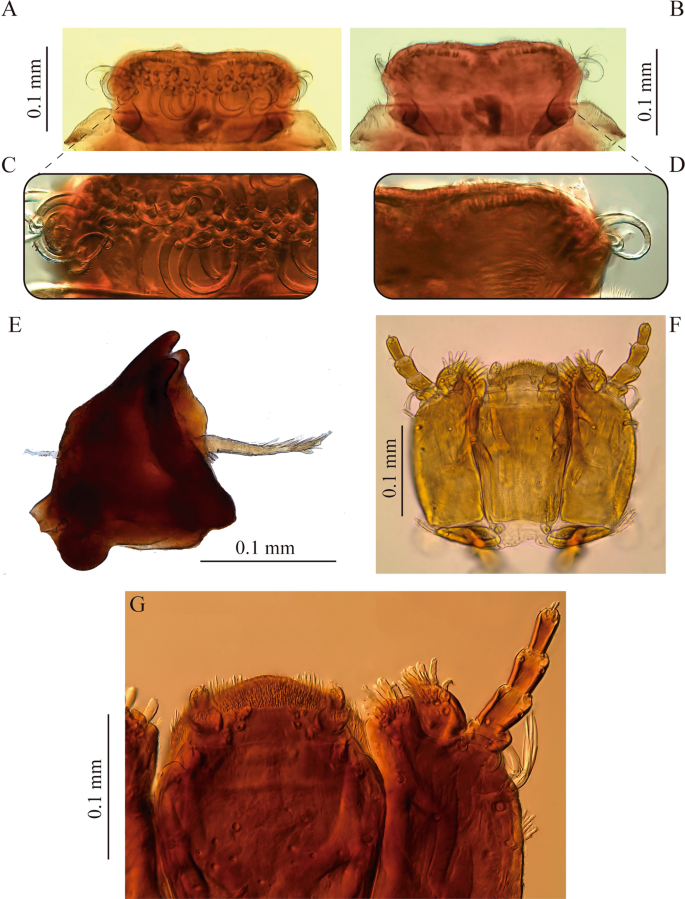
Mouthparts of S. rufulus larva. (A-D) labrum, (A) dorsal view, (B) ventral view, (C) dorsal detail, (D) ventral detail, (E) mandible, (F-G) maxillolabial complex.
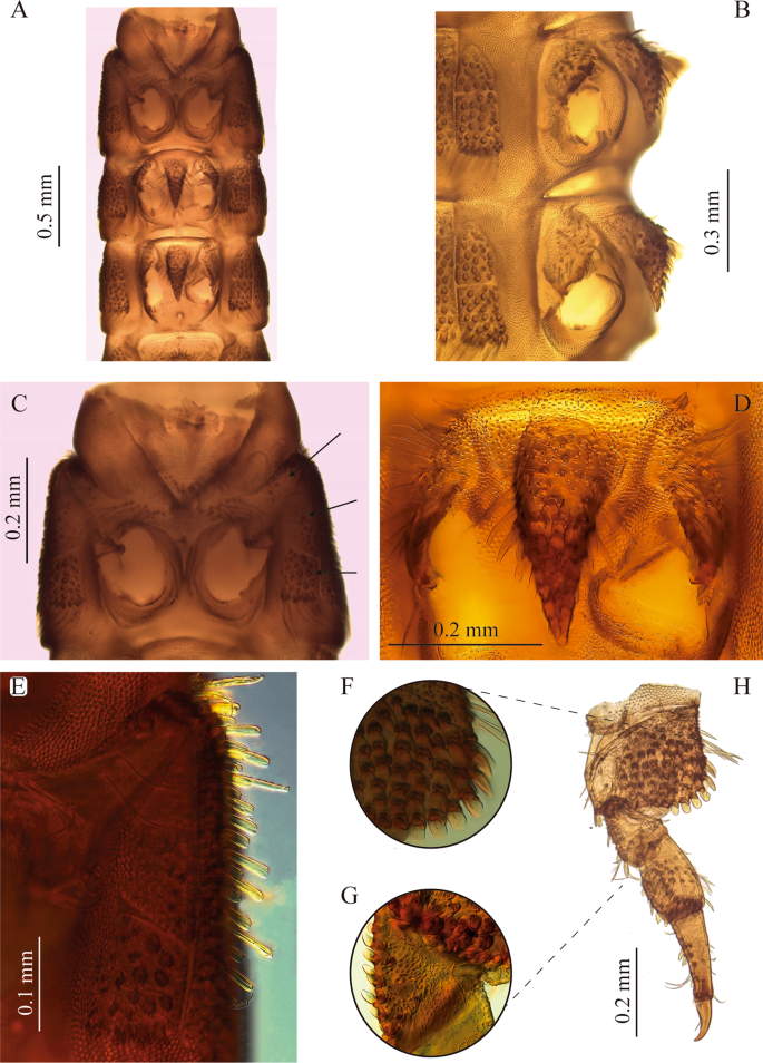
Thorax and legs of S. rufulus larva. (A) Thorax, ventral view, (B) meso- and metathorax, lateral view, (C) prosternum, (D) mesosternal process (E) prosternal sclerites, (F) coxa, (G) trochanter, (H) metathoracic leg. Arrows show prosternal sclerites.
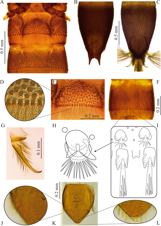
Abdomen of S. rufulus larva. (A) Abdominal sternites I – II, (B,C) abdominal segment IX, (B) dorsal, (C) ventral, (D,E) abdominal segment I, (D) sternum detail (E) posterior margin (F) abdominal segment IV, without sternal sutures in ventral view, (G) anal hook, (H) fan-shaped-tufted seta, (I) ramose setae, posterior margin of an abdominal segment, (J–L) anal operculum, (J) lateral margin, (K) external view, (L) apex.
Description of mature larva of Stenhelmoides rufulus (Hinton, 1934)
Body
Elongate, cylindrical, sides subparallel, thorax slightly wider than abdomen (Fig. 1). Color yellowish brown to reddish brown (Fig. 1). Tegument profusely tuberculate with several kinds of setae: tufted, either short or long, branched, spatulate, spine-like and scale-like (Figs. 2B, 5D,H–I). Anterior region of head, venter of thorax, legs and sternum of first abdominal segment remarkably covered by strong spatulate and scale-like setae. Length: 6.8–8.8 mm; maximum width: 0.87–1.05 mm.
Head
Capable of retraction into the thorax. Head capsule subquadrangular (Fig. 2A) (slightly wider than long). Dorsal surface at basal half smooth, except by some scattered punctures and two sublateral rows with four short setae lined on basal fourth (parietal area). Dorsal surface of anterior half coated with abundant, long, translucent, spatulate setae projected posteriad, more prominent on the sides. Coronal line long, about 1/4 as long as head capsule. Frontal lines long, extending to inner margin of antennal sockets. Frontoclypeal suture present. Frons protruding beyond antennae, with rounded anterolateral edges and anterior margin almost straight, anterior region without teeth. Ocular region on each side of head lighter in color and closer to base of antenna than to posterior end of head capsule. Lacking stemmata, but some individuals with one or two ocular pigment granules without lens.
Antenna
As long as half of head capsule (Fig. 2A,D). Basal antennomere (A1) short, slightly wider than long, bearing a crown of ramose setae. Second antennomere (A2) the longest, 1.5 times longer than first, bearing a distal long and prominent sensorium; slightly longer than second antennomere. Third antennomere (A3) the shortest, about 1/8 as long as second antennomere, bearing a tiny apical seta.
Mouthparts
Labrum subrectangular, convex, slightly wider at distal third (Fig. 3A,B). Anterior margin emarginated at middle, more evident from ventral view. Anterolateral angles rounded. Dorsal surface with a transverse band of strong long curled plumose setae arranged at about distal third. Ventral side with three stout simple setae on anterolateral corners, anterior margin with short simple setae with large sockets. Rest of ventral surface (epipharynx) covered by a dense pubescence oriented mesad and posteriad.
Mandibles
Strongly sclerotized, symmetrical, as long as wide, apex with three blunt teeth (Fig. 3E). Inner margin on dorsal side (dorsal carina) sharp, area between dorsal carina and mesal edge concave, with a brush of hair-like setae (pennicilus). Prostheca present, stout and plumose, shorter than mandible length, mesally oriented. Outer margin of dorsal surface with one tufted seta at about mid-length.
Maxilla
Basally with a transverse, narrow, suboval cardo, bearing a short tufted seta closer to outer margin (Fig. 3F,G). Stipes large, subrectangular, twice longer than wide; external (or ventral) surface of stipes with several setae distributed as follows: a longitudinal row of short tufted setae on outer margin, three short setae arranged as an oblique line from near base of lacinia to closer to outer margin and two long slender curled tufted setae near base of palpus. Lacinia and galea strongly sclerotized; lacinia subquadrangular, fully fused to stipes, apex truncated with several apical and subapical stout setae distally as well as on inner margin; galea entire, one-segmented, suboval, shorter than lacinia, bearing a group of stout distal setae. Palpus four-segmented, first palpomere the shortest with a curled seta; second palpomere twice as long as first, lacking setae; third palpomere slightly longer than second, with two apical setae, one on inner margin and one on outer margin; distal palpomere slightly shorter than second, bearing several short apical sensilla.
Labium
Well developed, formed by a large postmentum and a short prementum (Fig. 3F,G). Postmentum 1.5 times longer than wide, slightly narrower basally; anterolateral corners projected outwardly as lobes bearing a small digitiform sensillum; ventral surface with scattered pores clustered mainly on distal area of disc and two pairs of setae, one pair of short tufted setae at each distal corner and one pair on apical third, at each lateral margin; two small rounded sclerites near base of postmentum, bearing a long-stemmed tufted seta. Prementum short, wider than long, with a basal membranous region and an apical sclerotized plate bearing the palpi, disc with two spatulate setae and distal margin densely setose. Palpus two-segmented, palpiger present, well sclerotized; both palpomeres subequal in length; first palpomere bearing several short setae on outer apical corner; second palpomere narrower, bearing several apical sensilla.
Proventriculus
Equipped with strongly sclerotized teeth arranged as follows (Fig. 2F,G): one anterior row of clustered laminar teeth followed by a posterior band forming a well-defined belt of suboval teeth bearing about four tiny teeth; whole structure wider than long.
Thorax
Strongly sclerotized (Figs. 1D–G, 4); tergal plates with sagittal lines and densely covered with setiferous tubercles, except prothorax, which is smooth and shiny with tuberculate zones confined to lateral portions; posterior margin of all thoracic segments with a row of very long ramose setae, as long as adjacent membranous area. Ventral region of all thoracic segments with large reticulate membranous areas between sclerites. Prothorax the longest segment, subtrapezoidal, wider basally, slightly notched near apical margin, with a conspicuous anterior membranous neck, three times wider than long; dorsal surface sparsely and finely puncticulate, except for lateral areas which are densely covered with coarse setiferous tubercles; lateral margins with large spatulate setae and two basal tufted setae. Ventral region of prothorax with six sclerites, two antero-lateral subtriangular poorly sclerotized sclerites extending from lateral margin to near outer margin of coxal cavity, with long, slender tufted setae; two lateral pairs, the anteriormost subtriangular with conical and spatulate setae and membranous area continuing over inner margin of this sclerite, and the posteriormost subrectangular with coarse setiferous tubercles, conical and tufted setae. Posterior margin of ventral region of meso- and metathorax with a median circular impression. Meso- and metathorax wider than long, dorsally covered with setiferous tubercles and with an anterior ring of rugulose sculpture; ventrally with seven sclerites, one large anterior median conical process-like sclerite rising from a poorly sclerotized trapezoidal base, and two sub-median smaller subtriangular sclerites at each side of median sclerite, and two lateral pairs, one anterior subtriangular and one posterior subrectangular; coxal cavities open. Mesothorax bearing a pair of lateral spiracles.
Legs
Five-segmented, subequal in chaetotaxy and size (Fig. 4F–H). Coxa the largest segment, strong and subrectangular, external surface forming a protruding plate apically crenulated; plate with spine-like and scale-like setae on the disc and scale-like setae on edges; distal margin with a ring of scale-like setae; basal margin with scattered tufted setae, a patch of spine-like setae, few simple setae and basal spine-like sculpturing; apical edge emarginated in front of trochanter with scale-like setae; inner surface with a few simple and tufted setae on disc and apical scale-like and spine-like setae. Trochanter subtriangular, slightly shorter than femur, most of the surface covered by small spines and several scattered scale-like setae and a few ramose setae located mainly on the disc and edges; external surface with two spine-like setae on apical edge, near dorsal area. Femur subrectangular wider than tibia; anterior region almost glabrous with a row of long tufted setae at external edge; posterior margin coated with prominent setiferous tubercles and scale-like setae, internal surface with three spine-like setae and apical long tufted setae, external surface without setae or with some scattered pores. Tibia longer than femur, subrectangular to subtrapezoidal (base wider than the apex), anterior region with a row of short tufted setae and one medial long curly tufted setae, posterior region with two patches (subbasal and subapical) of tubercles with scale-like setae and two rows of scattered small and long tufted setae, external surface with two subbasal scale-like setae and posterior spine-like sculptures. Pretarsus subtriangular with a tiny distal seta slightly shorter than claw; claw blunt-pointed slightly curved (obtuse angle) at apical third.
Abdomen
Ninesegmented (Fig. 1A–C), strongly sclerotized. Surface covered by bicuspidated tubercles bearing a fan-shaped-tufted seta and a curly seta surrounding the tubercle, such as in thorax (Fig. 5D,H,I). Anterior edge of each abdominal segment with spine-like sculptures developing gradually from the membranous area to sclerotized area with larger tubercles. Posterior margins with a belt of quadrangular bicuspidated tubercles bearing a long ramose seta intercalated with a smaller tubercle accompanied by a shorter simple seta (Fig. 5D,I).
Segments I-VIII subequal in length, wider than long, segments VII-VIII almost as long as wide, segment IX the longest, 2 times longer than segment VIII with basal half parallel-sided and distal half tapering toward apex (Fig. 5B,C). Abdominal apex with a U-shaped emargination bearing thick tufted setae and two apical spinous processes 1/4 as long as segment IX (Fig. 5B). These projections are bare but surrounded by various setal types on base. Spiracles present on segments I-VIII. Segment I with complete sagittal line, segments II-IX lacking sagittal line. Pleural sclerite fusiform, only present on first abdominal segment, wider basally and narrowing towards the apex, inner margin (sternopleural suture) rounded and protruding above the sclerite, external margin (tergopleural suture) oblique on the same plane as the rest of the segment (Fig. 5A). Sternal plate semicircular, wider than long with strong scale-like and tufted setae on edges (Fig. 5E). Sternopleural and tergopleural sutures complete on first segment, incomplete on second segment reaching the anterior half (Fig. 5A). Segments III to IX without defined sutures, as complete rings with texture and tuberculation similar in ventral and dorsal face (Fig. 5F).
Operculum
Subpentagonal, longer than wide (Fig. 5K). Basal two thirds quadrate, lateral margins with short setae, external surface with two to three longitudinal rows of bicuspid tubercles on disc, rest of surface glabrous (Fig. 5K). Distal third acute, profusely hairy with tufted setae longer than those of lateral margins (Fig. 5J–L). Inner surface densely coated with simple setae, lateral vertex with thick tufted setae.
Anal hooks sharp on apical fifth obtusely curved close to 90° (Fig. 5G). Hooks non-serrated, lacking teeth on lateral margin. External and inner surfaces with long setae, slightly longer at inner base. Basal sclerite of hook twice as wide as hook base, bearing long setae. Gills composed by three tufts of simple gill filaments (more than 20 in each tuft).
Larval material examined
COLOMBIA • 1 specimen; Caquetá; Florencia; Morelia; El Dedo creek; Hacha basin river; 1°38′58.7″N, 75°38′34.7″W; 309 m a.s.l.; 3 feb. 2009; S. Gaspar; M. Peláez-Rodríguez legs; rock; MUSENUV • 1 specimen; Chocó; Quibdó; Tutunendo river; N 5°44′32″N, 76°31′17.22″W; 96 m a.s.l.; 21 sep. 2004; S. Sánchez leg; rock; CLCH • 2 specimens; Córdoba; Tierralta; Tuis-Tuis creek; 8°1′26.29″N, 76°4′53.18″W; 225 m a.s.l.; 24 oct. 2014; J. Garcés leg; MUSENUV • 1 specimen; La Guajira; San Juan del Cesar; Caracolí; Ranchería river; 10°57′13.6″N, 73°3′15.6″W; 465 m a.s.l.; 18 oct. 2009; C. Tamaris; G. Rúa; C. Guzmán legs; leaf litter; MUSENUV • 2 specimens; Magdalena; Aracataca; Tucurinca river; 10°41′16.3″N, 74°1′29.3″W; 500 m a.s.l.; 8 may. 2010; C. Tamaris; T. Sierra; G. Rúa legs; gravel; MUSENUV • 3 specimens; the same information except Ciénaga; Toribío river; 11°2′59.21″N, 74°13′3.79″W; 30 m a.s.l.; 9 jun. 2007; CMA-UCO code1438 • 1 specimen; Valle del Cauca; Buenaventura; La Nevera creek; Dagua basin river; 3°48′26″ N, 76°46′50″W; 360 m a.s.l.; 19 feb. 1999; M. del C. Zúñiga; S. Mosquera; G. Guevara legs; rock; MUSENUV.
Adult material of Stenhelmoides rufulus examined
COLOMBIA • 1♀; Caquetá; Florencia; before Florencia; Hacha river; 1°38′47.9″N, 75°36′37.1″W; 385 m a.s.l.; 30 sep. 2013; N. Oviedo leg; COMAC-Code 0589/Coleop 0135 • 1 specimen; Vereda Paraiso; Paraiso river; Hacha basin river; 71°38′58″N, 75°37′45″W; 682 m a.s.l.; 31 oct. 2013; S. Gaspar; M. Peláez-Rodríguez legs; rock; MUSENUV • 1♀; Chocó; Quibdó; Tutunendo river; 5°45′58.27″N, 76°33′33.23″W; 36 m a.s.l.; 18 oct. 2003. CMA-UCO Code1143 • 1 specimen; Córdoba; Tierralta; Tuis-Tuis creek (E1); 8°1′26.29″N, 76°4′53.18″W; 225 m a.s.l.; 24 oct. 2014; J. Garcés leg; MUSENUV • 2 specimens; the same information except (E2); 8°1′49.21″N, 76°5′11.30″W; 204 m a.s.l.; MUSENUV • 1♀; La Guajira; Cerrejón; 21 jun. 1981; Surber ned; IAvH-Code 140959 • 1♀, 1♀; Magdalena; Aracataca; Tucurinca; Tucurinca river; 10°41′16.3″N, 74°1′29.3″W; 500 m a.s.l.; 8 may. 2010; C. Tamaris; T. Sierra; G. Rúa legs; gravel; MUSENUV.
Habitat
Larvae and adults of Stenhelmoides were collected within rocky substrates and leafy litter situated in streams between 30 and 682 meters above sea level (m a.s.l.). The studied streams are located in three main natural geographic Colombian regions: the Pacific (two sites), the Caribbean (four sites), and the Andean portion along the foothill of the Amazon region (three sites) (Figs. 6 and 7A–D).
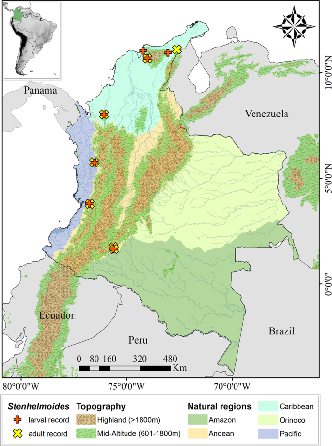
Map of the distribution of Stenhelmoides larval and adult records in this work.
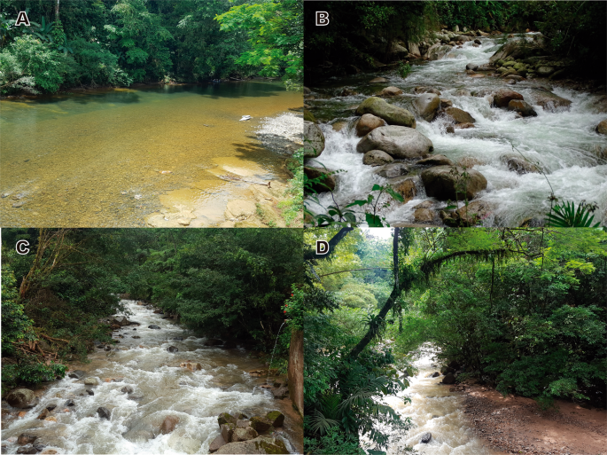
Sampling sites. (A) Tutunendo river, (B) Tucurinca river, (C) Paraiso River, (D) El Dedo creek. Photos: Zuleyma Mosquera (A); César Tamaris (B); Juliette Pauline Chaux (C,D).
The Pacific region is located in the western portion of Colombia. The streams within this region belong to the macro-basins of the Atrato and Dagua rivers in the Choco Biogeographic ecoregion. This zone is recognized for its high biodiversity and profuse endemism. The predominant life zones are the tropical rain forest (bp-T) and the very humid tropical forest (bmh-T). In addition, this zone has an extensive hydric network due to intense rainfall, with some places reporting a multi-year average precipitation as high as 10,000 mm29,30.
The Caribbean region is located in the northern portion of Colombia and South America. The largest number of larvae and adults of Stenhelmoides were collected in this region, with sampling sites located in the foothills of different fronts of the Sierra Nevada de Santa Marta (SNSM) and in lowlands of the Valley of the Sinú River. The SNSM offers a rich hydric network with streams characterized by short lengths and significant altitudinal gradients due to the steep slopes and confined basins in the area. The predominant vegetation is associated with the humid subtropical forest (bh-BT), mostly present in forests along poorly preserved riverbanks. The deforestation in the zone has been favored to promote agricultural activities, which has generated great pressure on the hydrographic basins31.
The Andean-Amazonian transition is located in the southern portion of Colombia and the collection sites correspond to the Department of Caquetá. The foothills of this mountain range have the sub-Andean forest as the primary vegetation class, which gradually merges with the upper layer of the high-Andean forest and the lower layer of the humid forest of warm floors. These ecosystems have special ecological conditions associated with the transitional environment between the fauna of Andean origin and the fauna of the Amazon plains. Despite having their own characteristics, the vegetation layers gradually mix due to the changes in the altitudinal gradients. The sub-Andean forest has been fragmented over the last decades and has been affected by agricultural activities, which has resulted in the loss of large areas of forest and the deterioration of the quality of the water bodies32.
The sampling events were carried out in a heterogeneous group of water currents with different flow rates (Fig. 7). Three of them correspond to creeks with flows considered of low-order magnitude, generally between 0.25 and 0.80 m3.sec−1, and with abundant areas of rapids and rocky beds. Other six sites are located in rivers with medium- to high-order magnitude flow rates. Specifically, the Tutunendo river is the water body with the highest flow rate in the study, reporting a medium 157.67 m3.sec−1, a minimum 57.58 m3.sec−1, and a maximum 257.58 m3.sec−1 monthly multi-year average flows. In addition, the Hacha river reported a medium 34.5 m3/sec, a minimum 19.33 m3.sec−1, and a maximum 156.92 m3.sec−1 monthly multi-year average flows, while the Rancheria river reported a medium 6.17 m3/sec, a minimum 3.24 m3.sec−1, and a maximum 35.71 m3.sec−1 monthly multi-year average flows33. Out of this group, the Tucurinca and Toribío rivers in the Sierra Nevada de Santa Marta are the water bodies with the lowest flow rate in the study, with point measurements between 1.5 to 2.1 m3.sec−1 at the sampling locations in the middle basins. The Paraiso river has no information available for the flow. These creeks and rivers belong to multiple hydrographic regions in Colombia that drain to the Atlantic and Pacific Oceans and the Amazon River macro-basin.
In general, the sampled streams had relatively clear fresh waters (19 to 22 °C), with dissolved oxygen levels close to saturation and with low residual organic loads. The perimeter areas of the riparian corridors exhibited a different type and magnitude of vegetation coverage, with a secondary forest condition in a relatively good conservation state. For those streams with available information, the water quality index values fluctuated between 50 and 74 units (ICA-FSN)34 which correspond to a good environmental quality category. The oxygen saturation levels between 87% and 100%, along with biochemical oxygen demands between 0.5 and 1.5 mg.LO2−1, indicate that organic contamination in the streams is low. The pH values between 6.8 and 7.9 units indicate the presence of a stable acid-buffer system, while the specific conductance levels between 25 and 140 μ.cm−1, reveal low mineralization of the waters in the studied streams31,35 (CINARA, unpublished data). Although the distribution and substrates of larvae and adults were similar for the specimens collected as part of this investigation, both stages do not seem to share the same microhabitat.
Associated Fauna
In this study, Stenhelmoides was found co-occurring with larvae and adults of Austrolimnius formosus (Sharp, 1882), Cylloepus Erichson, 1847, Disersus Sharp, 1882, Heterelmis Sharp, 1882, Hexanchorus Sharp, 1882, Huleechius Brown, 1981, Macrelmis Motschulsky, 1860, Microcylloepus Hinton, 1935, Neocylloepus chaparensis Manzo & Moya, 2010, Neoelmis Musgrave, 1935, Phanocerus congener Grouvelle, 1898, Pharceonus volcanus Spangler & Santiago-Fragoso, 1992 and Xenelmis Hinton, 1936.
Source: Ecology - nature.com

