Test organism
The dung beetle T. lusitanicus was selected as test species. This species has been successfully used in other physiological studies32,33 due to its easy rearing and the high volume of haemolymph in its body. Specimens of T. lusitanicus were used to study ivermectin biomagnification patterns from cattle dung into the gut, haemolymph, fat body and excreta (biological matrices) to gain insight about the pharmacokinetic behaviour of ivermectin in dung beetles.
In November 2016, individuals of T. lusitanicus were collected at La Sauceda, an ivermectin-free site in the Los Alcornocales Natural Park (Cádiz), in Southern Spain (36°31′54″N, 5°34′29″W). We used pitfall traps baited with cow dung to capture live beetles. The dung beetles were maintained in aerated plastic containers (80 × 30 × 50 cm) at 18 °C with 65% relative humidity (RH) and a photoperiod of 14:10 h (light:dark). The substrate was moss and dead fallen leaves of Quercus to prevent beetle stress.
In order to standardize the physiological condition of the beetles, only mature specimens were selected according to external age-grading methods (e.g., abrasion of the fore tibiae in conjunction with cuticle hardness of the pronotum and elytra, which makes it possible to sort out the individuals of approximately the same age34). In addition, we used a 1:1 sex ratio in each experiment. This work conforms to the Spanish legal requirements including those relating to conservation and welfare.
Biological matrices
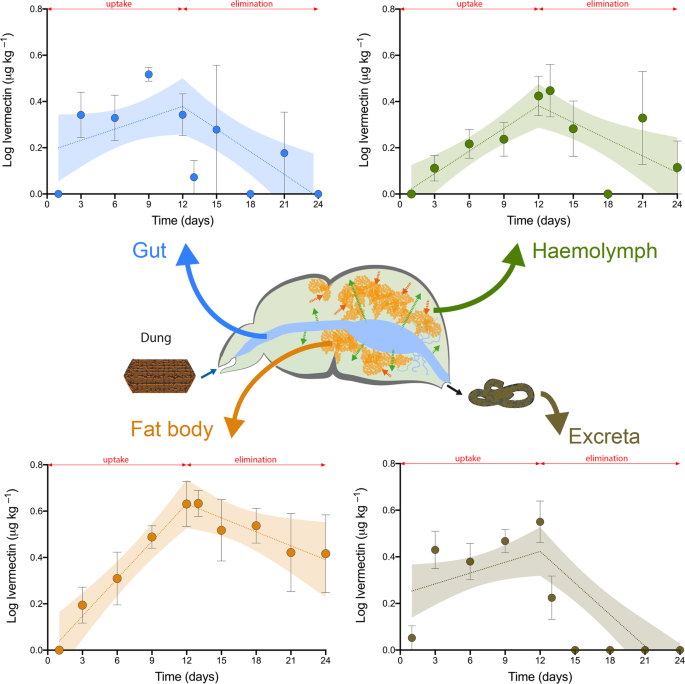
To determine the accumulation of ivermectin in dung beetles, four different matrices (gut, haemolymph, fat body, and excreta) were chosen. Ivermectin concentrations in the gut and excreta were analysed because they indicate the amount of ivermectin ingested and excreted, respectively. Haemolymph has been selected since it is the fluid in direct contact with all tissues and organs, and is responsible for the distribution of a large number of biological constituents to the cells (amino acids, proteins, lipids, and carbohydrates, among others). Fat body was selected because it constitutes the main tissue for nutrient storage, intermediary metabolism and regulation being the insect equivalent to the adipose tissue and liver in vertebrates28.
Application of the test substance and preparation of experimental diets
For this toxicological test, a concentration of 10 μg kg−1 (fresh weight) of ivermectin (≥90% B1a; ≤5% B1b percent purity, Sigma Aldrich Co., St. Louis, USA) in dung and an untreated dung control were used. The ivermectin concentration used was chosen because it is closely related to the toxicity threshold corresponding to the inhibition of antennal response in dung beetles (IC50 = 8.16 μg kg−1), as previously reported by Verdú et al.15. This concentration was also selected because it corresponds to the OECD guidelines, which suggest that the chosen concentration should be low enough to ensure that it does not affect insect behaviour while being high enough to allow its quantification throughout the uptake and elimination phases20. Dung treatments were prepared using the procedure developed by Verdú et al.13, in which a single concentration of ivermectin was spiked into fresh cow dung. The solution was made by dissolving ivermectin in absolute ethanol (Sigma-Aldrich Co.), subsequently added to 2 kg of fresh dung, and mixed for 2 h using a dough mixer machine. The concentration of ivermectin in the dung was determined after spiking to verify its homogenous distribution in the dung. The untreated dung control was processed in an identical manner but without the addition of ivermectin. Dung treatments were exposed to air for 4 h to remove residual ethanol and then placed in sealed plastic buckets to prevent desiccation during storage at 5 °C until use.
Toxicological test design
According to the OECD standard technical guide20, we implemented a continuous 12-day uptake phase. Dung beetles (n = 18 individuals per treatment) were sampled at 1, 3, 6, 9 and 12 days after the first addition of dung with ivermectin. After this 12-day uptake phase, the dung beetles were transferred to clean test boxes for the subsequent 12-day elimination phase also sampled at 1, 3, 6, 9 and 12 days. The experiments are described in the sections below.
Experimental procedure
During the continuous 12-day uptake phase, beetles were provided daily with 2 g of dung containing a concentration of 10 μg kg−1 (fresh weight) of ivermectin. After this 12-day uptake phase, the same individuals were provided with the same quantity (2 g) of ivermectin-free dung and were sampled again during another 12-days period (at 1, 3, 6, 9 and 12 days after the end of the first feeding phase). At each sampling time, food remains were removed, and their dry weight recorded to estimate the feeding rate per individual, which was standardised according to the fresh body weight of each individual (g day−1 g−1). During all the experimental period, dung beetles were maintained individually in plastic containers (60 × 40 × 40 cm) with moist sterile paper as a substrate at 18 °C (a temperature similar to the optimal temperature experienced in the field) within a climate-controlled chamber at 65% relative humidity (RH) and a photoperiod of 14:10 h (light:dark).
Validity of the test
In addition, to control the potential negative effects of ivermectin on beetle behaviour (pre-lethal symptoms), feeding rate, body weight, lipid content, and survival, 36 individuals were randomly assigned to two treatments: i) a control treatment (n = 18 individuals), in which beetles were fed with ivermectin-free dung during the whole 24 days experiment; and ii) an ivermectin treatment (n = 18 individuals), in which beetles were fed during 12 days (uptake phase) with spiked dung (containing ivermectin), followed by 12 days of feeding on ivermectin-free dung (elimination phase). Food consumption for each individual was monitored daily by placing 2 g of dung on a plastic dish (4 mm diameter and 13 mm height) to allow measurement of the quantity of dung ingested while avoiding water loss due to absorption by the substrate.
Sample preparation
To assess the biomagnification patterns and pharmacokinetic behaviour of ivermectin released from cattle dung, we extracted ivermectin from four biological matrices (gut, haemolymph, fat body and excreta) of T. lusitanicus. At the end of each of the ten sampling times (five during the uptake phase and five during the elimination phase), the gut, fat body, haemolymph and defecated beetle excreta were obtained from 10 randomly selected individuals. After each individual was anaesthetised with CO2, haemolymph samples were collected by puncturing the cuticle on the dorsal side of the pronotum and gently squeezing the beetle as described previously by Verdú et al.32. Haemolymph collected from each individual was placed into a vial, protected from light and maintained at –25 °C. Next, the beetle was placed on a dissection tray in which distilled water was added. The gut samples were obtained by dissecting from the foregut (stomodeum) to the hindgut (proctodeum) with the help of micro-clamps sealing both ends to avoid possible contamination. After dissection, the external surface was thoroughly washed with distilled water to rinse away any haemolymph that remained on the surface. Based on a previous work with the same test species32, to determine the lipid content, all of the fat body tissue was removed, placed on a dry paper towel to remove haemolymph and dried at 28 °C for 24 h to eliminate excess water prior to weighing using an AS 82/220.R2 analytical balance (RADWAG USA L.L.C., North Miami Beach, FL, USA). The samples of fat body and gut were taken from each individual and preserved in 300 µl of absolute ethanol. Before extraction process, the absolute ethanol from fat body and gut samples was evaporated with a gentle stream of nitrogen until dryness at room temperature. The excreta samples collected after defecation from each individual were dried and homogenised with liquid nitrogen and were then placed into plastic vials. To reach the minimum amount of material necessary to perform the analyses (0.3 ± 0.01 g for each sample) it was necessary to homogenise the samples from two individuals for each time period and biological matrix, with the exception of the gut on the third day of the uptake phase in which only a sample from a single individual was necessary. All samples were stored at −85 °C in a freezer (SANYO Electric Co. Ltd, Japan) until further analysis.
Ivermectin extraction
After the samples were collected, the clean up and extraction protocol was based on the optimised method described by Ortiz et al.35. Briefly, 0.3 ± 0.01 g of each sample (gut, haemolymph, fat body or excreta) was weighed in a 10 ml centrifuge tube, and 1 ml of a solution of acetonitrile:water (60:40) and 200 µl of the internal standard (IS) working solution (Abamectin, 10 ng ml−1) were added. The mixture was vortexed (REAX Control, Heidolph, Kelheim, Germany) during 1 min. After vortex mixing, the sample was sonicated using an ultrasonic homogenizer (SONOPLUS Ultrasonic Homogenizers HD 3200, 200 W, 20 KHz) at 40% power for 5 min. The tubes were centrifuged (BL-II, JP Selecta, Barcelona, Spain) at 5000 rpm for 10 min. The supernatant extracts were carefully removed, filtered (PTFE syringe filter 0.20 µm, Millex® Millipore Ibérica, Madrid, Spain) and transferred to a clean tube where it were evaporated to dryness at 45 °C under a stream of ultrahigh-purity N2. After reconstitution with 5 ml of pure water, the resulting solution was ultrasonicated using a water bath (JP Selecta, Barcelona, Spain) for 2 min and was filtered through a 0.45 µm PTFE syringe filter (Millex® Millipore Ibérica, Madrid, Spain) to remove any precipitate and to prevent the suspended particles from reaching the continuous unit.
The pre-concentration and clean-up of samples were performed using continuous solid phase extraction (SPE). Before pre-concentration of each sample, the SPE column was conditioned passing through the sorbent 1 ml of acetonitrile, 1 ml of methanol and 5 ml of purified water. The continuous SPE technique used was assembled from a Minipuls-3 peristaltic pump (Gilson, Middleton, WI, USA) fitted with polyvinyl chloride (PVC) pumping tubes, two Rheodyne 5041 injection valves (Cotati, CA, USA), PTFE tubing (3 mm I.D.) and a laboratory-prepared sorption column containing 80 mg Oasis-HLB® sorbent (5 cm × 3 mm i.d.). In the concentrating step, 5 ml of the reconstituted aqueous sample was passed at 4 ml min−1 through the sorbent column, thus the ivermectin and IS were adsorbed and the sample matrix was sent to waste. The analytes were eluted with 300 µl of acetonitrile into a glass vial and stored at −20 °C until analysis.
Reagents and chemicals
Standard grade ivermectin (≥90% B1a ≤5% B1b purity), and abamectin (≥98.7% purity) were purchased from Sigma-Aldrich (MO, USA) and stored at −20 °C. Abamectin, a precursor of ivermectin, differs from ivermectin in that it has a double bond at the C22–23 position34 and was used as an internal standard (chemical structures and physical chemical properties of ivermectin and abamectin are described in Supplementary Table S1 and Fig. S1). Oasis-HLB® sorbent was obtained from Waters Corporation (Milford, Massachusetts, USA). Ammonium formate and formic acid (analytical reagent grade) were purchased from Merck (Darmstadt, Germany). Millex-LG filter units (hydrophilic, PTFE, pore size = 0.20 and 0.45 μm, diameter = 25 mm, filtration area = 3.9 cm2) were obtained from Millipore Ibérica (Madrid, Spain). Water was purified by a Milli-Q purification system (Millipore Ibérica, Madrid, Spain). All other reagents used were purchased from standard suppliers and of analytical grade or higher.
Stock solutions
The method of standardization was based on the use of abamectin as internal standard, which was added to the samples just before the extraction procedure. Standard solutions were prepared as follows: to prepare the stock solutions, 1.0 ± 0.001 mg of ivermectin and abamectin reference standards were accurately weighed into individual 100 ml volumetric flasks and dissolved using methanol to prepare two stock solutions at a final concentration of 10 μg ml−1. Working solutions of ivermectin ranging from 0.1 to 10 ng ml−1 were prepared by appropriate dilution of the stock solution with methanol. The method of standardization was based on the use of abamectin as internal standard, which was added to the samples just before the extraction procedure. The internal standard working solution concentration was 10 ng ml−1. All of the solutions were stored in a freezer at −20 °C.
LC/ESI+–MS/MS instrumentation and settings
We applied the method proposed by Ortiz et al.35 for the quantitative determination of ivermectin. Briefly, ivermectin was analysed on an Agilent 1100 HPLC system, which was coupled to an Ion Trap MS analyser (Esquire 6000, Bruker Daltonics, Bremen, Germany) equipped with an electrospray ionization source (ESI). The system was controlled with the Agilent ChemStation (version A.06.01, Agilent Technologies) and Bruker Daltonics Esquire control (version 6.08, Bruker Daltonics) software packages. The data were processed with Data Analysis software (version 3.2, Bruker Daltonics).
A C18 Kinetex analytical column (75 mm × 3.0 mm, 3.0 μm, Phenomenex, Torrence, CA, USA) was used and it was operated at 40 °C. The mobile phase solvent A was a solution of 0.1% (v/v) formic acid in water, and solvent B was 0.1% (v/v) formic acid in acetonitrile. The column flow rate was 0.25 ml min−1, the injection volume was 1 μl and the gradient elution timetable was as follows: 0–10 min, 50% A-50% B; 10–15 min, 100% B; 15–20 min 50% A-50% B.
The MS/MS conditions were initially optimized by injecting standards at concentration of 1 μg/ml of ivermectin and internal standard solution. The optimal MS/MS sensitivity was obtained using electrospray in positive ionization (ESI+) and for quantitative purposes, the instrument was operated in multiple reaction monitoring (MRM) mode, scanning from 50 to 2000 m/z. In positive ion mode, a precursor ion at m/z 897 [M + Na]+ was observed for ivermectin, similar to results shown for IS at m/z 895 [M + Na]+. For ivermectin, a major product ion at m/z 753.1 [M-144+Na]+ was observed an identical adduct ion for abamectin at m/z 751.1. Since the [M-144+Na]+ was the product ion with the greatest abundance, it was chosen for quantitation of both compounds.
The method for the quantitative determination of ivermectin in the different biological matrices was validated by linearity of the method, lower limit of detection (LLOD), lower limit of quantification (LLOQ) and percentage of recovery35 (see Supplementary Table S2).
Data analysis and statistics
Following OECD guidelines for the testing of chemicals20, if a steady state is not achieved within the uptake phase, the dietary biomagnification factor (BMFk) must be determined as the ratio between the uptake (ks) and elimination (ke) rate constants. For each fraction, the kinetic parameters (ks and ke) were calculated in one run by applying a piecewise linear regression model after log transformation of the data from both the uptake and elimination phases, simultaneously. The breakpoint was 12 days coinciding with the time when the beetles have stopped eating dung containing ivermectin. Least squares regression fitting was used to obtain confidence intervals for slope parameters (95% CI) and to quantify the goodness-of-fit of the regression (R2). To discard any possible negative effects caused by ivermectin, the beetle body weight, lipid content and survival under laboratory conditions were compared between treated and control individuals by means of t tests. All data were analysed using the software GraphPad Prism (v8, San Diego, USA).
Distribution factors (DF) were calculated for each tissue fraction to illustrate the differences in ivermectin accumulation rates between the distinct biological matrices of the studied beetles. The proportional distribution of total ivermectin residues in each of the four matrices was thus calculated to evaluate their contribution to the total ivermectin accumulated in all biological matrices at the end of the uptake (day 12) and elimination phases (day 24).
Source: Ecology - nature.com

